

| The Harrison's Principles of Internal Medicine, 18e, Videos & Animations feature comprehensive demonstrations and tutorials covering essential clinical procedures. (Requires QuickTime) |  |
PART 10: Disorders of the Cardiovascular System
SECTION 5 Vascular Disease
e33 Atlas of Percutaneous Revascularization (View PDF)
Case 1: Chronic Total Occlusion
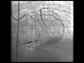 |
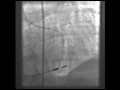
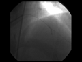
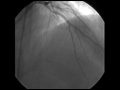
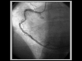
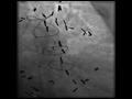
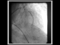
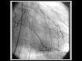
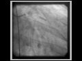
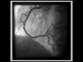
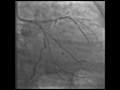
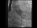
Video e33-1 Baseline left coronary angiogram shows an occluded LCx with left-to-left collaterals originating from LAD septal vessels. |
play video |
|
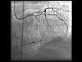 |
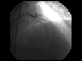
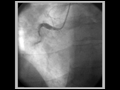
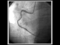
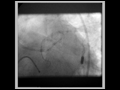
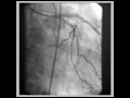
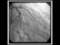
Video e33-2 Attempts to cross the total occlusion in the LCx using a hydrophilic wire and an antegrade approach were not successful, with the wire tracking to the right of the trajectory. |
play video |
|
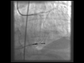 |
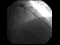
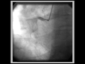
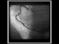
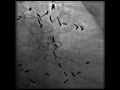
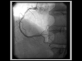
Video e33-3 The LAD septal collateral is accessed with a guidewire and directed toward the distal LCx to cross the total occlusion retrograde.
|
play video |
|
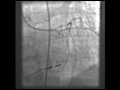 |
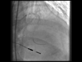
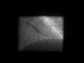
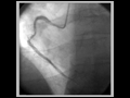
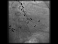
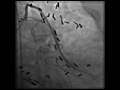
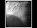
Video e33-4 The total occlusion is crossed retrograde. The wire is snared in the guide, exteriorized, and used to provide antegrade access to the LCx. |
play video |
|
 |
Video e33-5 Antegrade flow in the LCx is restored after balloon inflation. |
play video |
|
 |
Video e33-6 Following stenting of the total occlusion, blood flow in the distal vessel is improved and a second significant stenosis is seen. |
play video |
|
 |
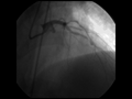
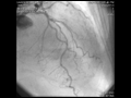
Video e33-7 Final result after LCx stenting. |
play video |
Case 2: Birfurcation Stenting
 |
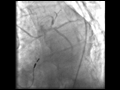
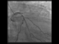
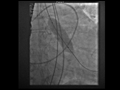
Video e33-8 Baseline angiogram of the left coronary circulation shows the significant stenosis in the mid-LAD and the bifurcation lesion involving a large diagonal branch. |
play video |
|
 |
Video e33-9 Both vessels are accessed with guidewires and pretreated with balloon angioplasty. |
play video |
|
 |
Video e33-10 Result after balloon angioplasty. |
play video |
|
 |
Video e33-11 Stent being positioned in the LAD. |
play video |
|
 |
Video e33-12 LAD post-stent result. |
play video |
|
 |
Video e33-13 Stent deployed in diagonal branch through the stent struts in the LAD using the "culotte" technique. |
play video |
|
 |
Video e33-14 Diagonal branch post-stent result. |
play video |
|
 |
Video e33-15 Simultaneous inflation of two 2.5-mm "kissing" balloons. |
play video |
|
 |
Video e33-16 Final postbifurcation stenting result. |
play video |
Case 3: Inferior Myocardial Infarction-Thrombus and Manual Thrombectomy
 |
Video e33-17 The right coronary artery (RCA) is totally occluded with filling defects in the vessel after contrast injection, indicating thrombus is present in the vessel. |
play video |
|
 |
Video e33-18 An angioplasty wire is threaded through the thrombotic lesion, but this does not restore blood flow to the distal vessel.
|
play video |
|
 |
Video e33-19 Result after manual thrombectomy and thrombus extraction. The "culprit" ruptured plaque and residual thrombus are now apparent in the vessel. |
play video |
|
 |
Video e33-20 After balloon angioplasty and stenting, thrombus is still present. |
play video |
|
 |
Video e33-21 After repeat manual thrombectomy and expansion of the stent, the thrombus is no longer present. |
play video |
|
 |
Video e33-22 Final Result. |
play video |
Case 4: Saphenous Vein Graft Intervention with Distal Protection
 |
Video e33-23 Saphenous vein graft to a first obtuse marginal branch with
an 80% eccentric stenosis in the midgraft. |
play video |
|
 |
Video e33-24 A distal protection device is deployed past the lesion. |
play video |
|
 |
Video e33-25 Angioplasty balloon inflation with the distal protection device in place. |
play video |
|
 |
Video e33-26 Final result after stent placement. |
play video |
Case 5: Unprotected Left Main PCI in a High-Risk Patient
 |
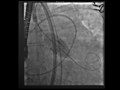
Video e33-27 Baseline left coronary artery injection in right anterior oblique (RAO) cranial projection shows a high-grade calcified stenosis in the left main coronary artery and a significant stenosis in the proximal LAD. |
play video |
|
 |
Video e33-28 In the left anterior oblique (LAO) caudal view, the left main coronary artery lesion can be seen to extend into the ostia of both the LCx and the LAD. |
play video |
|
 |
Video e33-29 Guide wires were placed into both the LCx and LAD. After the left main coronary artery and LCx are dilated with balloon angioplasty, the proximal LAD is dilated and a long drug-eluting stent is placed to... |
play video |
|
 |
Video e33-30 The bifurcation lesion in the left main coronary artery extending into the LCx and LAD ostia is treated using a "culotte" technique. First, a drug-eluting stent is placed in the left main coronary... |
play video |
|
 |
Video e33-31 Next, the LAD wire is removed and passed through the stent into the distal LAD. A second drug-eluting stent is deployed through the struts of the left main coronary artery/LCx stent. |
play video |
|
 |
Video e33-32 Following rewiring of the LCx, both stents are redilated simultaneously ("kissing" balloons). |
play video |
|
 |
Video e33-33 The final result in the LAO caudal view. |
play video |
|
 |
Video e33-34 The final result in the RAO cranial view showing patent left main, LCx, and LAD coronary arteries. |
play video |
Case 6: Multivessel PCI in a Diabetic Patient
 |
Video e33-35 Baseline angiogram of the left coronary circulation in the RAO view shows the total occlusion of the second obtuse marginal branch with delayed retrograde filling via collateral vessels and a high-grade... |
play video |
|
 |
Video e33-36 A guidewire is passed through the total occlusion and the lesion is pretreated with balloon angioplasty. |
play video |
|
 |
Video e33-37 Following placement of a drug-eluting stent in the lesion, the vessel is widely patent. A third obtuse marginal vessel, not previously seen, now fills faintly (TIMI 1 flow) with contrast but was not treated. |
play video |
|
 |
Video e33-38 The ramus intermedius lesion was crossed with a guidewire and pretreated with balloon angioplasty. |
play video |
|
 |
Video e33-39 A drug-eluting stent is placed across the ramus lesion and deployed. The final result shows no residual stenosis in either the ramus or second obtuse marginal vessels. |
play video |
|
 |
Video e33-40 Baseline angiogram of the RCA shows a high-grade lesion in the midsegment of the vessel. |
play video |
|
 |
Video e33-41 The lesion was pretreated with balloon dilation followed by stent deployment. |
play video |
|
 |
Video e33-42 The final result shows no residual stenosis in the mid-RCA. |
play video |
Case 7: Very Late Stent Thrombosis of a Proximal Lad Drug-Eluting Stent
 |
Video e33-43 Baseline angiogram showing a total occlusion of the proximal LAD within the drug-eluting stent and a significant stenosis at the origin of the LCx. |
play video |
|
 |
Video e33-44 The LAO view shows the LCx stenosis with a filling defect, indicating that thrombus is present in the vessel lumen. |
play video |
|
 |
Video e33-45 The LAD lesion was crossed with a guidewire, which resulted in slow filling of the mid-LAD (TIMI 2 flow), and revealed thrombus filling the stent. |
play video |
|
 |
Video e33-46 The final result after LAD and LCx stenting. The LAD lesion was pretreated with balloon angioplasty and a bare metal stent was deployed to cover the proximal lesion. The LCx ostial lesion was... |
play video |
Case 8: Transcatheter Aortic Valve Transplantation
 |
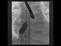
Video e33-47 Aortogram shows patent coronary arteries and minimal aortic insufficiency. |
play video |
|
 |
Video e33-48 Balloon valvuloplasty is performed with rapid ventricular pacing at 180 bpm. |
play video |
|
 |
Video e33-49 A 26-mm Edwards-SAPIEN valve is positioned using fluoroscopic and transesophageal echo guidance and deployed. |
play video |
|
 |
Video e33-50 Aortogram after valve deployment shows a functional valve with mild aortic insufficiency and without impingement of the coronary ostia. |
play video |
Case 9: Atrial Septal Defect Closure
 |
Video e33-51 A sizing balloon is placed across the ASD. |
play video |
|
 |
Video e33-52 An Amplatzer septal occluder is being positioned across the ASD. |
play video | |
 |
Video e33-53 The two discs of the device in place across the ASD. |
play video |