

| The Harrison's Principles of Internal Medicine, 18e, Videos & Animations feature comprehensive demonstrations and tutorials covering essential clinical procedures. (Requires QuickTime) |  |
PART 10: Disorders of the Cardiovascular System
SECTION 2 Diagnosis of Cardiovascular Disorders
e29 Atlas of Noninvasive Cardiac Imaging (View PDF)
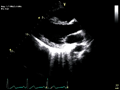 |
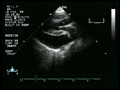
Video e29-1A Real-time two-dimensional echocardiographic images of a
patient with a normal heart. A. Parasternal long-axis view. There is symmetric contraction of the ventricles, evidenced by a decrease... |
play video |
 |
Video e29-1B Real-time two-dimensional echocardiographic images of a patient with a normal heart. B. Parasternal short-axis view. There is symmetric contraction of the ventricles, evidenced by a decrease... |
play video |
 |
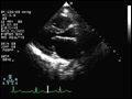
Video e29-2A Real-time two-dimensional echocardiographic images of a
patient with a severe decrease in left ventricular systolic function. The estimated ejection fraction is 20%. A. Parasternal long-axis view. |
play video |
 |
Video e29-2B Real-time two-dimensional echocardiographic images of a
patient with a severe decrease in left ventricular systolic function. The estimated ejection fraction is 20%. B. Parasternal short-axis view. |
play video |
 |
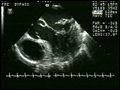
Video e29-3A Real-time two-dimensional echocardiographic images of a
patient with hypertrophic cardiomyopathy. There is a marked increase in left ventricular wall thickness with hyperdynamic systolic function...
|
play video |
 |
Video e29-3B Real-time two-dimensional echocardiographic images of a
patient with hypertrophic cardiomyopathy. There is a marked increase in left ventricular wall thickness with hyperdynamic systolic function...
|
play video |
 |
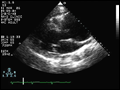
Video e29-4 Real-time two-dimensional parasternal long-axis images from a patient with aortic stenosis. There is normal left ventricular cavity size with normal systolic function. The aortic valve is thickened and... |
play video |
 |
Video e29-5A Real-time two-dimensional echocardiographic images of a
patient with mitral stenosis. There is diastolic doming and restricted leaflet opening secondary to fusion of the commissures. A. Parasternal... |
play video |
 |
Video e29-5B Real-time two-dimensional echocardiographic images of a
patient with mitral stenosis. There is diastolic doming and restricted leaflet opening secondary to fusion of the commissures. B. Parasternal... |
play video |
 |
Video e29-6A Real-time two-dimensional echocardiographic images from
the parasternal long-axis view of a patient with mitral valve prolapse. During systole, both anterior and posterior leaflet of the mitral valve... |
play video |
 |
Video e29-6B Real-time two-dimensional echocardiographic images from the parasternal long-axis view of a patient with mitral valve prolapse. During systole, both anterior and posterior leaflet of the mitral valve... |
play video |
 |
Video e29-7A Real-time two-dimensional images with color flow Doppler
imaging of a patient with mitral regurgitation due to ruptured chord tendineae. A. Gray-scale image showing a thickened redundant... |
play video |
 |
Video e29-7B Real-time two-dimensional images with color flow Doppler
imaging of a patient with mitral regurgitation due to ruptured chord tendineae. B. Color flow imaging showing severe mitral... |
play video |
 |
Video e29-8 Real-time transesophageal echocardiographic images of a
patient with severe mitral regurgitation due to a flail posterior leaflet. The posterior mitral valve leaflet is completely unsupported and moves into... |
play video |
 |
Video e29-9 Real-time two-dimensional echocardiographic images of a
patient with a vegetation on the mitral valve. There is a mobile echo density attached directly to the mitral valve apparatus that intermittently... |
play video |
 |
Video e29-10 Real-time transesophageal echocardiographic images of a
patient with a left atrial myxoma. There is a large echo-dense mass in the left atrium that is attached to the atrial septum. The mass moves across... |
play video |
 |
Video e29-11 Real-time two-dimensional echocardiographic images from the parasternal long-axis view of a patient with a large aneurysm of the ascending aorta. |
play video |
 |
Video e29-12 Real-time two-dimensional echocardiographic images of a
patient with pericardial effusion. The effusion is shown as a black echo-free space surrounding the heart. |
play video |
 |
Video e29-13 Real-time two-dimensional echocardiographic images from a subcostal view showing a large secundum atrial septal defect. There is a "drop out" in the region of the mid atrial septum. The right... |
play video |
 |
Video e29-14A Real-time two-dimensional echocardiographic images showing a close-up view of the atrial septum in a patient with the question of an atrial septal defect. A. Gray-scale image showing a questionable... |
play video |
 |
Video e29-14B Real-time two-dimensional echocardiographic images showing a close-up view of the atrial septum in a patient with the question of an atrial septal defect. B. Color flow imaging confirms... |
play video |
 |
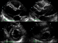
Video e29-15A Real-time two-dimensional stress echocardiogram in a
normal subject. The studies at rest are shown on the left and the studies during peak exercise are shown on the right. A. Parasternal long-axis... |
play video |
 |
Video e29-15B Real-time two-dimensional stress echocardiogram in a
normal subject. The studies at rest are shown on the left and the studies during peak exercise are shown on the right. B. Apical four-chamber... |
play video |
 |
Video e29-16A Real-time two-dimensional stress echocardiogram of a
patient with coronary artery disease. The studies at rest are shown on the left and studies during peak exercise are shown on the right... |
play video |
 |
Video e29-16B Real-time two-dimensional stress echocardiogram of a
patient with coronary artery disease. The studies at rest are shown on the left and studies during peak exercise are shown on the right... |
play video |
 |
Video e29-17 MRI scan in real time of a patient with a large left ventricular apical aneurysm. The long axis-view demonstrates a thin dyskinetic apical aneurysm with a preserved systolic function of the... |
play video |
 |
Video e29-18 Cine MRI scan of a patient with a dilated ascending aorta (annulo-aortic ectasia). There is a central jet of aortic regurgitation entering the left ventricular outflow tract. |
play video |
 |
Video e29-19 CT coronary angiogram showing a normal right coronary artery. The movie highlights multiple thin slices through the right coronary artery. |
play video |