

PART 19: High-Altitude and Decompression Sickness
e51 Altitude Illness
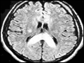 | Figure e51-1 T2 MRI image of the brain of a patient with HACE showing marked swelling and a hyperintense posterior body and splenium of the corpus callosum (area with dense opacity). The patient, a climber, went on to climb Mount Everest about 9 months after this episode of HACE. |
view large |
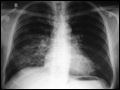 | Figure e51-2 Chest radiograph of a patient with HAPE shows opacity in the right mid- and lower zones simulating pneumonic consolidation. The opacity cleared almost completely in 2 days with descent and oxygen. |
view large |