

PART 17: Neurologic Disorders
SECTION 1 Diagnosis of Neurologic Disorders
e45 Electrodiagnostic Studies of Nervous System Disorders: EEG, Evoked Potentials, and EMG
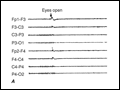 | Figure e45-1 A. Normal EEG showing a posteriorly situated 9-Hz alpha rhythm that attenuates with eye opening. B. Abnormal EEG showing irregular diffuse slow activity in an obtunded patient with encephalitis. |
view large |
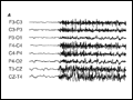 | Figure e45-2 Electrographic seizures.A. Onset of a tonic seizure showing generalized repetitive sharp activity with synchronous onset over both hemispheres. B. Burst of repetitive spikes occurring with sudden onset in the right... |
view large |
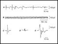 | Figure e45-3 Activity recorded during EMG.A. Spontaneous fibrillation potentials and positive sharp waves. B. Complex repetitive discharges recorded in partially denervated muscle at rest. |
view large |
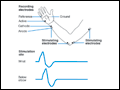 | Figure e45-4 Arrangement for motor conduction studies of the ulnar nerve. Responses are recorded with a surface electrode from the abductor digiti minimi muscle to supramaximal stimulation of the nerve at different sites, and are shown in the lower panel. (From Aminoff, 1998.) |
view large |