

PART 16: Endocrinology and Metabolism
SECTION 3 Disorders of Intermediary Metabolism
e41 Atlas of Clinical Manifestations of Metabolic Diseases
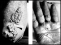
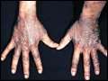 | Figure e41-1 "Gauntlet" of pellagra(niacin deficiency). Indurated, lichenified, pigmented, and scaly skin on the dorsa of the hands. (Source: K Wolff, RA Johnson, D Suurmond: Fitzpatrick's Color Atlas & Synopsis of Clinical Dermatology, 5th ed. New York, McGraw-Hill, 2005, www.accessmedicine.com.) See |
view large |
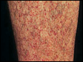 | Figure e41-2 Scurvy(vitamin C deficiency). Perifollicular hemorrhage on the leg. The follicles are often plugged by keratin (perifollicular hyperkeratosis). This eruption occurred in a 46-year-old alcoholic, homeless male who also had bleeding gums and loose teeth. (Source: K Wolff, RA... |
view large |
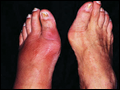 | Figure e41-3 Podagra with gouty inflammation of the first metatarsophalangeal (MTP) joint. Note the swelling and erythema of the left first MTP. (Courtesy of Kevin J. Knoop, MD, MS; with permission.) See Chaps. 333 and 359. |
view large |
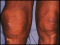 | Figure e41-4 Gout. Large tophi of gout located in and around the right knee. (Courtesy of Daniel L. Savitt, MD; with permission.) See Chaps. 333 and 359. |
view large |
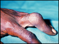 | Figure e41-5 Gout. The finger is an unusual site for gouty arthritis. Examination of the synovial fluid confirmed the diagnosis. (Courtesy of Alan B. Storrow, MD; with permission.) See Chaps. 333 and 359. |
view large |
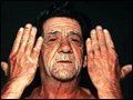
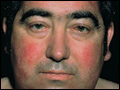 | Figure e41-6 Cushing's syndrome. Plethoric moon facies with erythema and telangiectases of cheek and forehead; the face and neck and supraclavicular areas (not depicted here) show increased deposition of fat. (Source: K Wolff, RA Johnson, D Suurmond: Fitzpatrick's Color Atlas & Synopsis of Clinical Dermatology, 5th... |
view large |
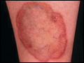 | Figure e41-7 Necrobiosis lipoidica diabeticorum. A large, symmetric plaque with active tan-pink, well-demarcated, raised, firm borders and a yellow center in the pretibial regions of a 28-year-old diabetic female. The central parts of the lesions are depressed with atrophic changes of epidermal thinning and telangiectasis against yellow... |
view large |
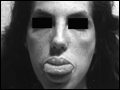 | Figure e41-8 Patient with multiple endocrine neoplasia 2B syndrome. Note the multiple neuromas on the lips and tongue and the marfanoid facies. (Source: DG Gardner, D Shoback, eds: Greenspan's Basic & Clinical Endocrinology, 8th ed. New York, McGraw-Hill, 2006, www.accessmedicine.com.) See |
view large |
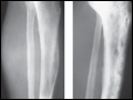 | Figure e41-9 Early and late radiographs of Paget's disease of the tibia, taken when the male patient was 45 years of age (A) and when he was 65 years of age (B). (Source: HB Skinner: Current Diagnosis & Treatment in Orthopedics, 4th ed. New York, McGraw-Hill, 2007, www.accessmedicine.com.) See |
view large |
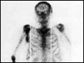 | Figure e41-10 Bone scan of patient with severe Paget's disease of the skull, ribs, spine, pelvis, right femur, and acetabulum. Note localization of bone-seeking isotope (99mTc-labeled bisphosphonate) in these areas. (Source: DG Gardner, D Shoback, eds: Greenspan's Basic & Clinical Endocrinology, 8th ed. New... |
view large |
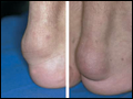 | Figure e41-11 Tendinous xanthomas. Large subcutaneous tumors adherent to the Achilles tendons. (Source: K Wolff, RA Johnson, D Suurmond: Fitzpatrick's Color Atlas & Synopsis of Clinical Dermatology, 5th ed. New York, McGraw-Hill, 2005, www.accessmedicine.com.) See |
view large |
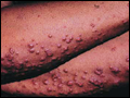 | Figure e41-12 Papular eruptive xanthomas. A. Multiple, discrete, red-to-yellow papules becoming confluent on the elbow of an individual with uncontrolled diabetes mellitus; lesions were present on both elbows and buttocks. B. Papular eruptive xanthomas on the... |
view large |
 | Figure e41-13 Forms of xanthomas and other lipid deposits frequently seen in familial hypercholesterolemia homozygotes.A. Arcus corneae. B, C, E, and F. Cutaneous planar... |
view large |
 | Figure e41-14 Examples of xanthomas in type III hyperlipoproteinemic subjects.A. Tuberoeruptive xanthomas of the elbows. B. Tuberous xanthomas of the digits and xanthomas of the palmar creases (xanthoma striata palmaris) (arrows). (Courtesy of Dr. Thomas P. Bersot; with permission.) See |
view large |
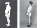
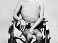 | Figure e41-15 A 17-year-old patient with abetalipoproteinemia with generalized weakness, kyphoscoliosis, and lordosis. (Courtesy of Drs. Peter Herbert, Gerd Assmann, Antonio M. Gotto, Jr., and Donald Fredrickson; with permission.) See |
view large |
 | Figure e41-16 Porphyria cutanea tarda. Periorbital and malar violaceous coloration, hyperpigmentation, and hypertrichosis on the face; bullae, crusts, and scars on the dorsa of the hands. (Source: K Wolff, RA Johnson, D Suurmond: Fitzpatrick's Color Atlas & Synopsis of Clinical Dermatology, 5th ed. New York,... |
view large |
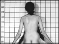
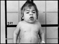 | Figure e41-17 Mucopolysaccharidosis type IH (Hurler's syndrome) in a 4-year-old boy. Diagnosis was made at the age of 15 months, at which time he had developmental delay, hepatomegaly, and skeletal involvement. At the time of the picture, the patient had short stature, an enlarged tongue, persistent nasal discharge, stiff joints, and... |
view large |
 | Figure e41-18 Growth and development in two patients with type Ia glycogen storage disease.A. Patient at age 7 years and at age 39 years. B. Another type Ia patient at age 10 years, and follow-up at age 33 years. Both patients survive... |
view large |
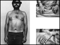 | Figure e41-19 Progressive myopathy in a patient with type IIIa glycogen storage disease. The patient has a debrancher deficiency in both liver and muscle (subtype IIIa). As a child, he had hepatomegaly, hypoglycemia, and growth retardation. After puberty, he no longer had hepatomegaly, and his final height is normal. Note the muscle wasting... |
view large |
 | Figure e41-20 Skeletal features of Marfan's syndrome in a 16-year-old girl. Note the long limbs that are associated with disproportionate tall stature, long fingers, scoliosis, and genu valgum. (Source: CR Scriver, AL Beaudet, WS Sly, D Valle, eds: The Metabolic and Molecular Bases of Inherited Disease online, 8th ed.... |
view large |
 | Figure e41-21 Marfan's syndrome.A. Long, narrow face. B. Arachnodactyly and positive wrist sign. C. High-arched palate. D.... |
view large |
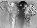 | Figure e41-22 Ochronotic pigmentation of the femur of a 56-year-old alkaptonuric patient. (Courtesy of Dr. H. W. Edmonds of the Washington Hospital Center, Washington, DC; with permission.) See |
view large |
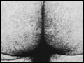 | Figure e41-23 Clusters of dark-red to blue angiokeratomas (telangiectases) on the buttocks (A) and in the umbilical area (B) of a hemizygote with Fabry disease. (Source: CR Scriver, AL Beaudet, WS Sly, D Valle, eds: The Metabolic and Molecular Bases of Inherited Disease online, 8th ed. New York, McGraw-Hill, www.ommbid.com.) See |
view large |
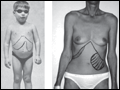 | Figure e41-24 Two patients with type B Niemann-Pick disease (NPD).A. A 4.7-year-old patient with type B NPD. (From DS Fredrickson, HR Sloan, in JB Stanbury et al: The Metabolic Basis of Inherited Disease, 3rd ed. New York, McGraw-Hill, 1972. Used by permission.) B. A 44-year-old patient with type B NPD. See |
view large |
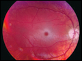 | Figure e41-25 "Cherry red" spot in the eye of a Tay-Sachs patient. See |
view large |
 | Figure e41-26 Kayser-Fleischer ring. This develops in Wilson's disease from copper deposition in Descemet's membrane, producing brownish discoloration of the peripheral cornea. It should not be confused with the yellow-white lipid ring of arcus senilis, which is common in the elderly and occasionally signifies hyperlipidemia, especially when... |
view large |
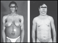 | Figure e41-27 Anterior view of patients with different forms of lipodystrophy.A. Congenital generalized lipodystrophy: a 16-year-old girl with generalized loss of fat, acromegaloid features, severe acanthosis nigricans affecting axillae and abdomen, umbilical hernia. ( |
view large |