

PART 2: Cardinal Manifestations and Presentation of Diseases
SECTION 9 Alterations in the Skin
e16 Atlas of Skin Manifestations of Internal Disease
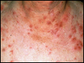
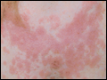
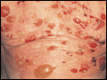
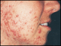 | Figure e16-1 Acne vulgaris with inflammatory papules, pustules, and comedones. (Courtesy of Kalman Watsky, MD; with permission.) |
view large |
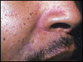
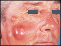
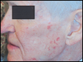
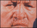
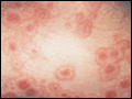
 | Figure e16-2 Acne rosacea with prominent facial erythema, telangiectasias, scattered papules, and small pustules. (Courtesy of Robert Swerlick, MD; with permission.) |
view large |
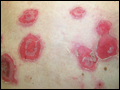
 | Figure e16-3 Psoriasis is characterized by small and large erythematous plaques with adherent silvery scale. |
view large |
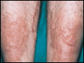
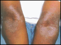 | Figure e16-4 Atopic dermatitis with hyperpigmentation, lichenification, and scaling in the antecubital fossae. (Courtesy of Robert Swerlick, MD; with permission.) |
view large |
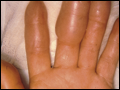
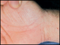
 | Figure e16-5 Dyshidrotic eczema, characterized by deep-seated vesicles and scaling on palms and lateral fingers, is often associated with an atopic diathesis. |
view large |
 | Figure e16-6 Seborrheic dermatitis showing erythema and scale in the nasolabial fold (Courtesy of Robert A. Swerlick, MD; with permission.) |
view large |
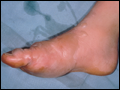
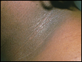
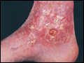 | Figure e16-7 Stasis dermatitis showing erythematous, scaly, and oozing patches over the lower leg. Several stasis ulcers are also seen in this patient. |
view large |
 | Figure e16-8 A. Allergic contact dermatitis, acute phase, with sharply demarcated, weeping, eczematous plaques in a perioral distribution. B. Allergic contact dermatitis to nickel, chronic phase demonstrating an erythematous, lichenified, weeping plaque on skin chronically exposed to a metal snap. (B, Courtesy of Robert Swerlick, MD; with... |
view large |
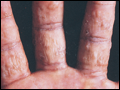
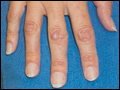
 | Figure e16-9 Lichen planus showing multiple flat-topped, violaceous papules and plaques. Nail dystrophy as seen in this patient's thumbnail may also be a feature. (Courtesy of Robert Swerlick, MD; with permission.) |
view large |
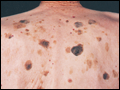 | Figure e16-10 Seborrheic keratoses are seen as “stuck on,” waxy, verrucous papules and plaques with a variety of colors ranging from light tan to black. |
view large |
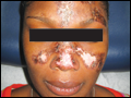
 | Figure e16-11 Vitiligo in a typical acral distribution demonstrating striking cutaneous depigmentation, as a result of loss of melanocytes. |
view large |
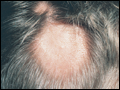 | Figure e16-12 Alopecia areata characterized by a sharply demarcated circular patch of scalp completely devoid of hairs. Follicular orifices are preserved, indicating a nonscarring alopecia. (Courtesy of Robert Swerlick, MD; with permission.) |
view large |
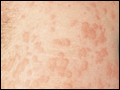
 | Figure e16-13 Pityriasis rosea. Multiple round to oval erythematous patches with fine central scale are distributed along the skin tension lines on the trunk. |
view large |
 | Figure e16-14 A. Urticaria showing characteristic discrete and confluent, edematous, erythematous papules and plaques. B. Dermatographism. Erythema and whealing that developed after firm stroking of the skin. (B, Courtesy of Robert Swerlick, MD; with... |
view large |
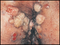
 | Figure e16-15 Epidermoid cysts. Several inflamed and noninflamed firm, cystic nodules are seen in this patient. Often a patulous follicular punctum is observed on the overlying epidermal surface. |
view large |
 | Figure e16-16 Keloids resulting from ear piercing, with firm exophytic flesh-colored to erythematous nodules of scar tissue. |
view large |
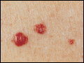 | Figure e16-17 Cherry hemangiomas are very common and arise in middle-aged to older adults. They are characterized by multiple erythematous to dark-purple papules, usually located on the trunk. |
view large |
 | Figure e16-18 Frostbite with vesiculation, surrounded by edema and erythema. (Courtesy of Daniel F. Danzl, MD; with permission.) |
view large |
 | Figure e16-19 Frostbite with vesiculation, surrounded by edema and erythema. (Courtesy of Daniel F. Danzl, MD; with permission.) |
view large |
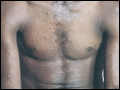
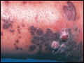 | Figure e16-20 Kaposi's sarcoma in a patient with AIDS demonstrating patch, plaque, and tumor stages. |
view large |
 | Figure e16-21 Non-Hodgkin's lymphoma involving the skin with typical violaceous, “plum-colored” nodules. (Courtesy of Jean Bolognia, MD; with permission.) |
view large |
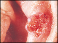 | Figure e16-22 Basal cell carcinoma showing central ulceration and a pearly, rolled, telangiectatic tumor border. |
view large |
 | Figure e16-23 Mycosis fungoides is a cutaneous T cell lymphoma, and plaque stage lesions are seen in this patient. |
view large |
 | Figure e16-24 Metastatic carcinoma to the skin is characterized by inflammatory, often ulcerated dermal nodules. |
view large |
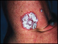
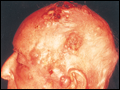
 | Figure e16-25 Keratoacanthoma is a low-grade squamous cell carcinoma that presents as an exophytic nodule with central keratinous debris. |
view large |
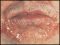
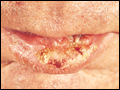 | Figure e16-26 Squamous cell carcinoma seen here as a hyperkeratotic crusted and somewhat eroded plaque on the lower lip. Sun-exposed skin such as the head, neck, hands, and arms are other typical sites of involvement. |
view large |
 | Figure e16-27 Actinic keratoses consist of hyperkeratotic erythematous papules and patches on sun-exposed skin. They arise in middle-aged to older adults and have some potential for malignant transformation. (Courtesy of Robert Swerlick, MD; with permission.) |
view large |
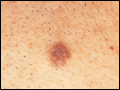 | Figure e16-28 Nevus. Nevi are benign proliferations of nevomelanocytes characterized by regularly shaped hyperpigmented macules or papules of a uniform color. |
view large |
 | Figure e16-29 Dysplastic nevi are irregularly pigmented and shaped nevomelanocytic lesions that may be associated with familial melanoma. |
view large |
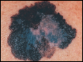 | Figure e16-30 Superficial spreading melanoma is the most common type of malignant melanoma and demonstrates color variegation (black, blue, brown, pink, and white) and irregular borders. |
view large |
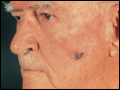 | Figure e16-31 Lentigo maligna melanoma occurs on sun-exposed skin as a large, hyperpigmented macule or plaque with irregular borders and variable pigmentation. (Courtesy of Alvin Solomon, MD; with permission.) |
view large |
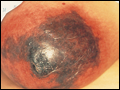
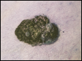 | Figure e16-32 Nodular melanoma most commonly manifests itself as a rapidly growing, often ulcerated or crusted black nodule. (Courtesy of S. Wright Caughman, MD; with permission.) |
view large |
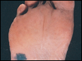 | Figure e16-33 Acral lentiginous melanoma is more common in blacks, Asians, and Hispanics and occurs as an enlarging hyperpigmented macule or plaque on the palms or soles. Lateral pigment diffusion is present. |
view large |
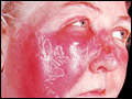 | Figure e16-34 Erysipelas is a streptococcal infection of the superficial dermis and consists of well-demarcated, erythematous, edematous, warm plaques. |
view large |
 | Figure e16-35 Varicella showing numerous lesions in various stages of evolution: vesicles on an erythematous base, umbilicated vesicles, and crusts. (Courtesy of Robert Hartman, MD; with permission.) |
view large |
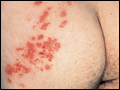 | Figure e16-36 Herpes zoster is seen in this HIV-infected patient as hemorrhagic vesicles and pustules on an erythematous base grouped in a dermatomal distribution. (Courtesy of Robert Swerlick, MD; with permission.) |
view large |
 | Figure e16-37 Impetigo contagiosa is a superficial streptococcal or Staphylococcus aureus infection consisting of honey-colored crusts and erythematous weeping erosions. Occasionally, bullous lesions may be seen. |
view large |
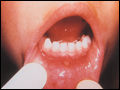 | Figure e16-38 Tender vesicles and erosions in the mouth of a patient with hand-foot-and-mouth disease. (Courtesy of Stephen D. Gellis, MD; with permission.) |
view large |
 | Figure e16-39 Lacy reticular rash of erythema infectiosum (fifth disease). |
view large |
 | Figure e16-40 Molluscum contagiosum is a cutaneous poxvirus infection characterized by multiple umbilicated flesh-colored or hypopigmented papules. (Courtesy of Yale Resident's Slide Collection; with permission.) |
view large |
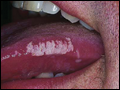 | Figure e16-41 Oral hairy leukoplakia often presents as white plaques on the lateral tongue and is associated with Epstein-Barr virus infection. (From K Wolff, RA Johnson, D Suurmond: Fitzpatrick's Color Atlas & Synopsis of Clinical Dermatology, 5th ed. New York, McGraw-Hill, 2005. |
view large |
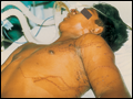
 | Figure e16-42 Fulminant meningococcemia with extensive angular purpuric patches. (Courtesy of Stephen D. Gellis, MD; with permission.) |
view large |
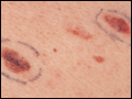
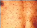
 | Figure e16-43 Rocky Mountain spotted fever demonstrating pinpoint petechial lesions on the palm and volar aspect of the wrist. (Courtesy of Robert Swerlick, MD; with permission.) |
view large |
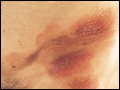
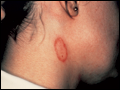
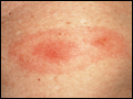 | Figure e16-44 Erythema chronicum migrans is the early cutaneous manifestation of Lyme disease and is characterized by erythematous annular patches, often with a central erythematous papule at the tick bite site. (Courtesy of Yale Resident's Slide Collection; with permission.) |
view large |
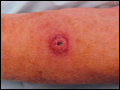
 | Figure e16-45 Primary syphilis with a firm, nontender chancre. (Courtesy of Gregory Cox, MD; with permission.) |
view large |
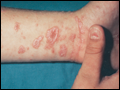
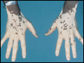
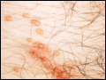
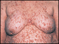
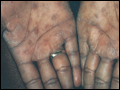 | Figure e16-46 Secondary syphilis commonly affects the palms and soles with scaling, firm, red-brown papules. (Courtesy of Alvin Solomon, MD; with permission.) |
view large |
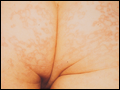
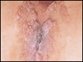
 | Figure e16-47 Condylomata lata are moist, somewhat verrucous intertriginous plaques seen in secondary syphilis. (Courtesy of Yale Resident's Slide Collection; with permission.) |
view large |
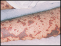
 | Figure e16-48 Secondary syphilis demonstrating the papulosquamous truncal eruption. |
view large |
 | Figure e16-49 Tinea corporis is a superficial fungal infection, seen here as an erythematous annular scaly plaque with central clearing. |
view large |
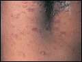
 | Figure e16-50 Scabies showing typical scaling erythematous papules and few linear burrows. |
view large |
 | Figure e16-51 Skin lesions caused by Chironex fleckeri sting. (Courtesy of V. Pranava Murthy, MD; with permission.) |
view large |
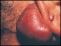
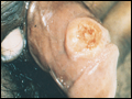
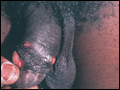 | Figure e16-52 Chancroid with characteristic penile ulcers and associated left inguinal adenitis (bubo). |
view large |
 | Figure e16-53 Condylomata acuminata are lesions induced by human papillomavirus and in this patient are seen as multiple verrucous papules coalescing into plaques. (Courtesy of S. Wright Caughman, MD; with permission.) |
view large |
 | Figure e16-54 A patient with features of polar lepromatous leprosy; multiple nodular skin lesions, particularly of the forehead, and loss of eyebrows. (Courtesy of Robert Gelber, MD; with permission.) |
view large |
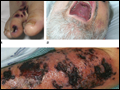 | Figure e16-55 Skin lesions of neutropenic patients. A. Hemorrhagic papules on the foot in a patient undergoing treatment for multiple myeloma. Biopsy and culture demonstrated Aspergillosis sp. B. Eroded nodule on the hard palate of a... |
view large |
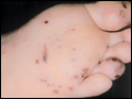 | Figure e16-56 Septic emboli with hemorrhage and infarction due to acute Staphylococcus aureus endocarditis. (Courtesy of L. Baden, MD; with permission.) |
view large |
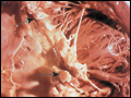 | Figure e16-57 Vegetations (arrows) due to viridans streptococcal endocarditis involving the mitral valve (Courtesy of AW Kerchner, MD; with permission) |
view large |
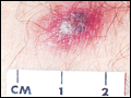 | Figure e16-58 Disseminated gonococcemia in the skin is seen as hemorrhagic papules and pustules with purpuric centers in an acral distribution. (Courtesy of Daniel M. Musher, MD; with permission.) |
view large |
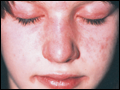
 | Figure e16-59 A. Systemic lupus erythematosus showing prominent, scaly, malar erythema. Involvement of other sun-exposed sites is also common. B. Acute lupus erythematosus on the upper chest demonstrating brightly erythematous and slightly edematous coalescence of papules and plaques. (B, Courtesy of Robert Swerlick, MD; with... |
view large |
 | Figure e16-60 Discoid lupus erythematosus. Atrophic, depigmented plaques and patches surrounded by hyperpigmentation and erythema associated with scarring and alopecia, are characteristic of this cutaneous form of lupus. |
view large |
 | Figure e16-61 Dermatomyositis. Periorbital violaceous erythema characterizes the classic heliotrope rash. (Courtesy of James Krell, MD; with permission.) |
view large |
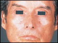 | Figure e16-62 Scleroderma characterized by typical expressionless, mask-like facies. |
view large |
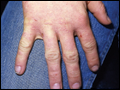
 | Figure e16-63 Dermatomyositis often involves the hands as erythematous flat-topped papules over the knuckles (Gottron's sign) and periungual telangiectasias. |
view large |
 | Figure e16-64 Scleroderma showing acral sclerosis and focal digital ulcers. |
view large |
 | Figure e16-65 Erythema multiforme is characterized by multiple erythematous plaques with a target or iris morphology and usually represents a hypersensitivity reaction to drugs or infections (especially herpes simplex virus). (Courtesy of Yale Resident's Slide Collection; with permission.) |
view large |
 | Figure e16-66 Dermatitis herpetiformis manifested by pruritic, grouped vesicles in a typical location. The vesicles are often excoriated and may occur on knees, buttocks, elbows and posterior scalp. |
view large |
 | Figure e16-67 A. Pemphigus vulgaris demonstrating eroded bullae on the back. B. Pemphigus vulgaris almost invariably involves the oral mucosa and may present with erosions involving the gingiva, buccal mucosa, palate, posterior pharynx, or the tongue. (B, Courtesy of Robert Swerlick, MD; with... |
view large |
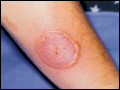
 | Figure e16-68 Erythema nodosum is a panniculitis characterized by tender deep-seated nodules and plaques usually located on the lower extremities. (Courtesy of Robert Swerlick, MD; with permission.) |
view large |
 | Figure e16-69 Vasculitis. Palpable purpuric papules on the lower legs are seen in this patient with cutaneous small vessel vasculitis. (Courtesy of Robert Swerlick, MD; with permission.) |
view large |
 | Figure e16-70 Bullous pemphigoid with tense vesicles and bullae on an erythematous, urticarial base. (Courtesy of Yale Resident's Slide Collection; with permission.) |
view large |
 | Figure e16-71 Acanthosis nigricans demonstrating typical hyperpigmented axillary plaques with a velvet-like, verrucous surface on the neck. |
view large |
 | Figure e16-72 Pretibial myxedema manifesting as waxy, infiltrated plaques in a patient with Graves' disease. |
view large |
 | Figure e16-73 Plaque of Sweet's syndrome demonstrating an erythematous indurated plaque with a pseudo-vesicular border. (Courtesy of Robert Swerlick, MD, with permission.) |
view large |
 | Figure e16-74 Bilateral rheumatoid nodules of the upper extremities. (Courtesy of Robert Swerlick, MD; with permission.) |
view large |
 | Figure e16-75 Neurofibromatosis demonstrating numerous flesh-colored cutaneous neurofibromas. |
view large |
 | Figure e16-76 Coumarin necrosis showing cutaneous and subcutaneous necrosis of a breast. Other fatty areas such as buttocks and thighs are also common sites of involvement. (Courtesy of Kim Yancey, MD; with permission.) |
view large |
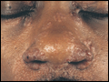 | Figure e16-77 A. Sarcoid. Infiltrated papules and plaques of variable color are seen in a typical paranasal and periorbital location. B. Sarcoid. Infiltrated, hyperpigmented, and slightly erythematous coalescent papules and plaques on the upper arm. (B, Courtesy of Robert Swerlick, MD; with... |
view large |
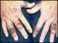 | Figure e16-78 Pyoderma gangrenosum on the dorsal aspect of both hands demonstrating multiple necrotic ulcers surrounded by a violaceous and undermined border. (Courtesy of Robert Swerlick, MD; with permission.) |
view large |