

PART 17: Neurologic Disorders
SECTION 3 Nerve and Muscle Disorders
387 Muscular Dystrophies and Other Muscle Diseases
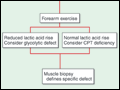 | Figure 387-1 Diagnostic evaluation of intermittent weakness. AChR AB, acetylcholine receptor antibody; CPT, carnitine palmitoyltransferase; EOMs, extraocular muscles; MG, myasthenia gravis; PP, periodic paralysis. |
view large |
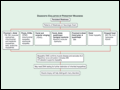 | Figure 387-2 Diagnostic evaluation of persistent weakness. Examination reveals one of seven patterns of weakness. The pattern of weakness in combination with the laboratory evaluation leads to a diagnosis. EOMs, extraocular muscles; OPMD, oculopharyngeal muscular dystrophy; FSHD, facioscapulohumeral dystrophy; IBM, inclusion body myositis;... |
view large |
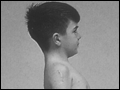
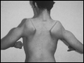 | Figure 387-3 Facioscapulohumeral dystrophy with prominent scapular winging. |
view large |
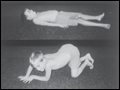 | Figure 387-4 Gowers' sign showing a patient using arms to climb up the legs in attempting to get up from the floor. |
view large |
 | Figure 387-5 Lordotic posture, exaggerated by standing on toes, associated with trunk and hip weakness. |
view large |
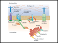 | Figure 387-6 Selected muscular dystrophy–associated proteins in the cell membrane and Golgi complex. |
view large |
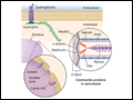 | Figure 387-7 Selected muscular dystrophy–associated proteins in the nuclear membrane and sarcomere. As shown in the exploded view, emerin and lamin A/C are constituents of the inner nuclear membrane. Several dystrophy-associated proteins are represented in the sarcomere including titin, nebulin, calpain, telethonin, actinin, and... |
view large |
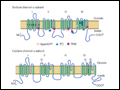 | Figure 387-8 The sodium and calcium channels are depicted here as containing four homologous domains, each with six membrane-spanning segments. The fourth segment of each domain bears positive charges and acts as the “voltage sensor” for the channel. The association of the four domains is thought to form a pore through which ions... |
view large |