

PART 17: Neurologic Disorders
SECTION 3 Nerve and Muscle Disorders
384 Peripheral Neuropathy
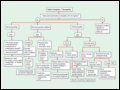 | Figure 384-1 Approach to the evaluation of peripheral neuropathies. CIDP, chronic inflammatory demyelinating polyradiculoneuropathy; GBS, Guillain-Barré syndrome. |
view large |
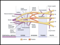 | Figure 384-2 Brachial plexus anatomy. L, lateral; M, medial; P, posterior. (From J Goodgold: Anatomical Correlates of Clinical Electromyography. Baltimore, Williams and Wilkins, 1974, p. 126, with permission.) |
view large |
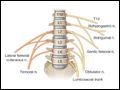 | Figure 384-3 Lumbar plexus. Posterior divisions are in orange, anterior divisions are in yellow. (From J Goodgold: Anatomical Correlates of Clinical Electromyography. Baltimore, Williams and Wilkins, 1974, p. 126, with permission.) |
view large |
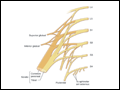 | Figure 384-4 Lumbosacral plexus. Posterior divisions are in orange, anterior divisions are in yellow. (From J Goodgold: Anatomical Correlates of Clinical Electromyography. Baltimore, Williams and Wilkins, 1974, p. 126, with permission.) |
view large |