

PART 17: Neurologic Disorders
SECTION 2 Diseases of the Central Nervous System
381 Meningitis, Encephalitis, Brain Abscess, and Empyema
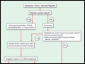 | Figure 381-1 The management of patients with suspected CNS infection. ADEM, acute disseminated encephalomyelitis; AFB, acid-fast bacillus; Ag, antigen; CSF, cerebrospinal fluid; CT, computed tomography; CTFV, Colorado tick fever virus; CXR, chest x-ray; DFA, direct fluorescent antibody; EBV,... |
view large |
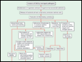 | Figure 381-2 The pathophysiology of the neurologic complications of bacterial meningitis. CSF, cerebrospinal fluid; SAS, subarachnoid space. |
view large |
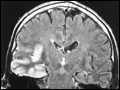 | Figure 381-3 Coronal FLAIR magnetic resonance image from a patient with herpes simplex encephalitis. Note the area of increased signal in the right temporal lobe (left side of image) confined predominantly to the gray matter. This patient had predominantly unilateral disease;... |
view large |
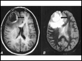 | Figure 381-4 Pneumococcal brain abscess. Note that the abscess wall has hyperintense signal on the axial T1-weighted MRI (A, black arrow), hypointense signal on the axial proton density images (B, black arrow), and enhances... |
view large |
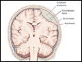 | Figure 381-5 Subdural empyema. |
view large |
 | Figure 381-6 Subdural empyema. There is marked enhancement of the dura and leptomeninges (A, B, straight arrows) along the left medial hemisphere. The pus is... |
view large |
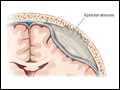 | Figure 381-7 Cranial epidural abscess is a collection of pus between the dura and the inner table of the skull. |
view large |
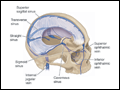 | Figure 381-8 Anatomy of the cerebral venous sinuses. |
view large |