

PART 17: Neurologic Disorders
SECTION 2 Diseases of the Central Nervous System
379 Primary and Metastatic Tumors of the Nervous System
 | Figure 379-1 Genetic and chromosomal alterations involved in the development of primary and secondary glioblastomas. A slash indicates one or the other or both. DCC, deleted in colorectal carcinoma; EGFR, epidermal growth... |
view large |
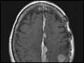
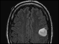 | Figure 379-2 Fluid-attenuated inversion recovery (FLAIR) MRI of a left frontal low-grade astrocytoma. This lesion did not enhance. |
view large |
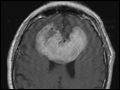
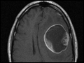 | Figure 379-3 Postgadolinium T1 MRI of a large cystic left frontal glioblastoma. |
view large |
 | Figure 379-4 Postgadolinium T1 MRI of a recurrent glioblastoma before (A) and after (B) administration of bevacizumab. Note the decreased enhancement and mass effect. |
view large |
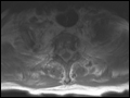
 | Figure 379-5 Postgadolinium T1 MRI demonstrating a large bifrontal primary central nervous system lymphoma (PCNSL). The periventricular location and diffuse enhancement pattern are characteristic of lymphoma. |
view large |
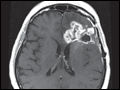
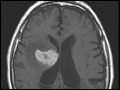
 | Figure 379-6 Postgadolinium T1 MRI demonstrating multiple meningiomas along the falx and left parietal cortex. |
view large |
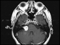 | Figure 379-7 Postgadolinium MRI of a right vestibular schwannoma. The tumor can be seen to involve the internal auditory canal. |
view large |
 | Figure 379-8 Postgadolinium T1 MRI of multiple brain metastases from non-small cell lung cancer involving the right frontal (A) and right cerebellar (B) hemispheres. Note the diffuse enhancement pattern and absence of central necrosis. |
view large |
 | Figure 379-9 Postgadolinium MRI images of extensive leptomeningeal metastases from breast cancer. Nodules along the dorsal surface of the spinal cord (A) and cauda equina (B) are seen. |
view large |
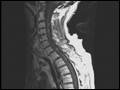
 | Figure 379-10 Postgadolinium T1 MRI showing circumferential epidural tumor around the thoracic spinal cord from esophageal cancer. |
view large |