

PART 17: Neurologic Disorders
SECTION 2 Diseases of the Central Nervous System
378 Concussion and Other Head Injuries
 | Figure 378-1 Traumatic cerebral contusion. Noncontrast CT scan demonstrating a hyperdense hemorrhagic region in the anterior temporal lobe. |
view large |
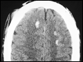 | Figure 378-2 Multiple small areas of hemorrhage and tissue disruption in the white matter of the frontal lobes on noncontrast CT scan. These appear to reflect an extreme type of the diffuse axonal shearing lesions that occur with closed head injury. |
view large |
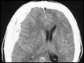 | Figure 378-3 Acute subdural hematoma. Noncontrast CT scan reveals a hyperdense clot which has an irregular border with the brain and causes more horizontal displacement (mass effect) than might be expected from its thickness. The disproportionate mass effect is the result of the large rostral-caudal... |
view large |
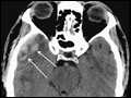
 | Figure 378-4 Acute epidural hematoma. The tightly attached dura is stripped from the inner table of the skull, producing a characteristic lenticular-shaped hemorrhage on noncontrast CT scan. Epidural hematomas are usually caused by tearing of the middle meningeal artery following fracture of the... |
view large |
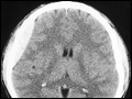
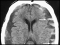 | Figure 378-5 CT scan of chronic bilateral subdural hematomas of different ages. The collections began as acute hematomas and have become hypodense in comparison to the adjacent brain after a period during which they were isodense and difficult to appreciate. Some areas of resolving blood are contained... |
view large |