

PART 17: Neurologic Disorders
SECTION 2 Diseases of the Central Nervous System
377 Diseases of the Spinal Cord
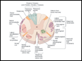 | Figure 377-1 Transverse section through the spinal cord, composite representation, illustrating the principal ascending (left) and descending (right) pathways. The lateral and ventral spinothalamic tracts (dark blue)... |
view large |
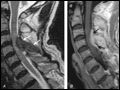
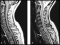 | Figure 377-2 Epidural spinal cord compression due to breast carcinoma. Sagittal T1-weighted (A) and T2-weighted (B) MRI scans through the cervicothoracic junction reveal an infiltrated and... |
view large |
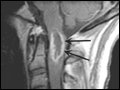
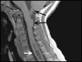
 | Figure 377-3 MRI of a thoracic meningioma. Coronal T1-weighted postcontrast image through the thoracic spinal cord demonstrates intense and uniform enhancement of a well-circumscribed extramedullary mass (arrows), which displaces the spinal cord to the left. |
view large |
 | Figure 377-4 MRI of an intramedullary astrocytoma. Sagittal T1-weighted postcontrast image through the cervical spine demonstrates expansion of the upper cervical spine by a mass lesion emanating from within the spinal cord at the cervicomedullary junction. Irregular peripheral enhancement occurs... |
view large |
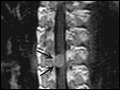
 | Figure 377-5 MRI of a spinal epidural abscess due to tuberculosis.A. Sagittal T2-weighted free spin-echo MR sequence. A hypointense mass replaces the posterior elements of C3 and extends epidurally to compress the spinal cord ( |
view large |
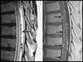 | Figure 377-6 Arteriovenous malformation. Sagittal MR scans of the thoracic spinal cord: T2 fast spin-echo technique (left) and T1 postcontrast image (right). On the T2-weighted image ( |
view large |
 | Figure 377-7 MRI of syringomyelia associated with a Chiari malformation. Sagittal T1-weighted image through the cervical and upper thoracic spine demonstrates descent of the cerebellar tonsils and vermis below the level of the foramen magnum (black arrows). Within the... |
view large |