

PART 17: Neurologic Disorders
SECTION 2 Diseases of the Central Nervous System
376 Trigeminal Neuralgia, Bell's Palsy, and Other Cranial Nerve Disorders
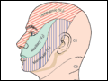 | Figure 376-1 The three major sensory divisions of the trigeminal nerve consist of the ophthalmic, maxillary, and mandibular nerves. |
view large |
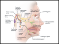 | Figure 376-2 The facial nerve. A, B, and C denote lesions of the facial nerve at the stylomastoid foramen, distal and proximal to the geniculate ganglion, respectively. Green lines indicate the parasympathetic fibers, red line indicates motor fibers, and purple lines indicate visceral afferent fibers... |
view large |
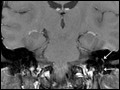 | Figure 376-3 Axial and coronal T1-weighted images post-Gadolinium with fat suppression demonstrate diffuse smooth linear enhancement of the left facial nerve, involving the genu, tympanic, and mastoid segments within the temporal bone (arrows), without evidence of mass... |
view large |
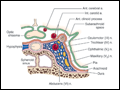 | Figure 376-4 Anatomy of the cavernous sinus in coronal section, illustrating the location of the cranial nerves in relation to the vascular sinus, internal carotid artery (which loops anteriorly to the section), and surrounding structures. |
view large |