

PART 17: Neurologic Disorders
SECTION 2 Diseases of the Central Nervous System
372 Parkinson's Disease and Other Movement Disorders
 | Figure 372-1 Pathologic specimens from a patient with Parkinson's disease (PD) compared to a normal control demonstrating (A) reduction of pigment in SNc in PD vs control, (B) reduced numbers of cells in SNc in PD compared to control, and (C) Lewy bodies (arrows) within melanized dopamine neurons in PD. SNc, substantia nigra pars... |
view large |
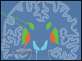 | Figure 372-2 Basal ganglia nuclei. Schematic (A) and postmortem (B) coronal sections illustrating the various components of the basal ganglia. SNc, substantia nigra pars compacta; STN, subthalamic nucleus. |
view large |
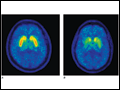
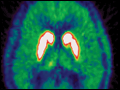 | Figure 372-3 Fluorodopa-PET in a normal individual (A) and a PD patient (B). Striatal FD-PET provides a measure of the integrity of the nigrostriatal system. Note reduced striatal uptake in PD compared to a control, which tends to be more pronounced in the caudate than in the putamen. (Courtesy of Dr. Jon... |
view large |
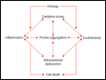 | Figure 372-4 Schematic representation of how pathogenetic factors implicated in PD interact in a network manner, ultimately leading to cell death. This figure illustrates how interference with any one of these factors may not necessarily stop the cell death cascade. (Adapted from CW Olanow: Movement Disorders, 22:S-335, 2007.) |
view large |
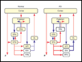 | Figure 372-5 Basal ganglia organization. Classic model of the organization of the basal ganglia in the normal, PD, and levodopa-induced dyskinesia state. Inhibitory connections are shown as blue arrows and excitatory connections as red arrows. The striatum is the major input region and receives its major input from the cortex. The GPi... |
view large |
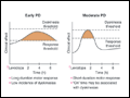 | Figure 372-6 Changes in motor response associated with chronic levodopa treatment. Levodopa-induced motor complications. Schematic illustration of the gradual shortening of the duration of a beneficial motor response to levodopa (wearing off) and the appearance of dyskinesias complicating “on” time. |
view large |
 | Figure 372-7 Treatment options for the management of PD. Decision points include: a. Introduction of a neuroprotective therapy: No drug has been established to have or is currently approved for neuroprotection or disease modification, but there are several agents that have this potential based on laboratory and preliminary clinical... |
view large |
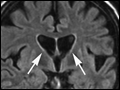 | Figure 372-8 Huntington's disease. A. Coronal FLAIR MRI shows enlargement of the lateral ventricles reflecting typical atrophy (arrows). B. Axial FLAIR image demonstrates abnormal high signal in the caudate and putamen (arrows). |
view large |