

PART 17: Neurologic Disorders
SECTION 2 Diseases of the Central Nervous System
371 Dementia
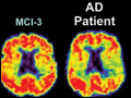 | Figure 371-1 PET images obtained with the amyloid-imaging agent Pittsburgh Compound-B ([11C]PIB) in a normal control (left); three different patients with mild cognitive impairment (MCI, center); and a mild AD patient (right). Some MCI patients have control-like... |
view large |
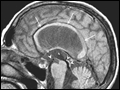
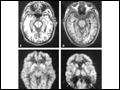 | Figure 371-2 Alzheimer's disease. Axial T1-weighted MR images through the midbrain of a normal 86-year-old athlete (A) and a 77-year-old man with AD (B). Note that both individuals have mild sulcal widening and slight dilation of the temporal horns of the lateral ventricles. However,... |
view large |
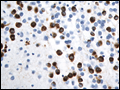
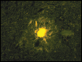 | Figure 371-3 Mature neuritic plaque with a dense central amyloid core surrounded by dystrophic neurites (thioflavin S stain). (Image courtesy of S DeArmond, University of California; with permission.) |
view large |
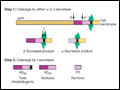 | Figure 371-4 Amyloid precursor protein (APP) is catabolized byα, β, andγsecretases. A key initial step is the digestion by either β-secretase (BASE) or α secretase [ADAM10 or ADAM17 (TACE)], producing smaller nontoxic products. Cleavage of the β... |
view large |
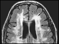 | Figure 371-5 Diffuse white matter disease. Axial fluid-attenuated inversion recovery (FLAIR) MR image through the lateral ventricles reveals multiple areas of hyperintensity involving the periventricular white matter as well as the corona radiata and striatum (arrows). While seen in some individuals with normal... |
view large |
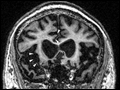 | Figure 371-6 Frontotemporal dementia (FTD). Coronal MRI sections from representative patients with behavioral variant FTD (left), semantic dementia (center), and progressive nonfluent aphasia (right). Areas of early and severe atrophy in each syndrome are... |
view large |
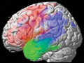 | Figure 371-7 Voxel-based morphometry analysis showing differing patterns of brain atrophy in three variants of progressive aphasia, including nonfluent (red), semantic (green), and logopedic subtypes (blue). Voxel-based morphometry allows comparison of MRI gray... |
view large |
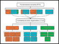 | Figure 371-8 Frontotemporal dementia syndromes are united by underlying frontotemporal lobar degeneration pathology, which can be divided according to the presence of tau, TPD-43, or fused in sarcoma (FUS) inclusions in neurons and glia. Correlations between clinical syndrome and major molecular category are shown with colored shading. |
view large |
 | Figure 371-9 Pick's disease, a subtype offrontotemporal lobar degeneration(FTLD)-tau. Pick bodies, shown here in the dentate gyrus of a patient with advanced bvFTD, consist of loosely arranged paired helical and straight filaments and stain positively for hyperphosphorylated tau.... |
view large |
 | Figure 371-10 Normal-pressure hydrocephalus. A. Sagittal T1-weighted MR image demonstrates dilation of the lateral ventricle and stretching of the corpus callosum (arrows), depression of the floor of the third ventricle (single arrowhead), and enlargement of... |
view large |