

PART 17: Neurologic Disorders
SECTION 1 Diagnosis of Neurologic Disorders
368 Neuroimaging in Neurologic Disorders
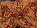 | Figure 368-1 CT angiography (CTA) of ruptured anterior cerebral artery aneurysm in a patient presenting with acute headache. A. Noncontrast CT demonstrates subarachnoid hemorrhage and mild obstructive hydrocephalus. B. Axial... |
view large |
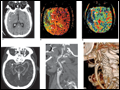 | Figure 368-2 Acute left hemiparesis due to middle cerebral artery occlusion. A. Axial noncontrast CT scan demonstrates high density within the right middle cerebral artery (arrow) associated with subtle low density involving the right... |
view large |
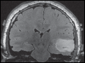
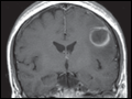 | Figure 368-3 Cerebral abscess in a patient with fever and a right hemiparesis. A. Coronal postcontrast T1-weighted image demonstrates a ring enhancing mass in the left frontal lobe. B. Axial diffusion-weighted image demonstrates restricted diffusion (high signal intensity) within the lesion, which in this setting is highly suggestive of cerebral... |
view large |
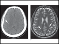
 | Figure 368-4 Herpes simplex encephalitis in a patient presenting with altered mental status and fever. A. and B. Coronal (A) and axial |
view large |
 | Figure 368-5 Susceptibility weighted imaging in a patient with familial cavernous malformations. A. Noncontrast CT scan shows one hyperdense lesion in the right hemisphere (arrow). B. T2-weighted fast spin echo image shows subtle... |
view large |
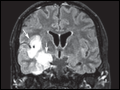
 | Figure 368-6 Diffusion tractography in cerebral glioma. A. An axial postcontrast T1-weighted image shows a nonenhancing glioma (T) of the left temporal lobe cortex lateral to the fibers of the internal capsule. B. Coronal T2 FLAIR image demonstrates high signal... |
view large |