

PART 17: Neurologic Disorders
SECTION 1 Diagnosis of Neurologic Disorders
366 Biology of Neurologic Diseases
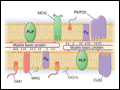 | Figure 366-1 The molecular architecture of the myelin sheath illustrating the most important disease-related proteins. The illustration represents a composite of CNS and PNS myelin. Proteins restricted to CNS myelin are shown in green, proteins of PNS myelin are lavender, and proteins present in both... |
view large |
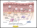 | Figure 366-2 A model for experimental allergic encephalomyelitis (EAE). Crucial steps for disease initiation and progression include peripheral activation of preexisting autoreactive T cells; homing to the CNS and extravasation across the blood-brain barrier; reactivation of T cells by exposed... |
view large |
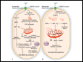 | Figure 366-3 Involvement of mitochondria in cell death. A severe excitotoxic insult (A) results in cell death by necrosis, whereas a mild excitotoxic insult (B) results in apoptosis. After a... |
view large |
 | Figure 366-4 Mirror neuron systems are bilaterally activated during imitation. A. Bilateral activations (circled in yellow) in inferior frontal mirror neuron areas during imitation, as measured by BOLD fMRI signal changes. In red, activation during right hand imitation. In... |
view large |