

PART 16: Endocrinology and Metabolism
SECTION 3 Disorders of Intermediary Metabolism
358 The Porphyrias
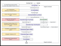 | Figure 358-1 The human heme biosynthetic pathway indicating in linked boxes the enzyme that, when deficient, causes the respective porphyria. Hepatic porphyrias are shown in yellow boxes and erythropoietic porphyrias in pink boxes. |
view large |
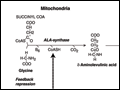 | Figure 358-2 The heme biosynthetic pathway showing the eight enzymes and their substrates and products. Four of the enzymes are localized in the mitochondria and four in the cytosol. |
view large |
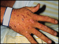 | Figure 358-3 Typical cutaneous lesions in a patient with porphyria cutanea tarda. Chronic, crusted lesions resulting from blistering due to photosensitivity on the dorsum of the hand of a PCT patient. (Courtesy of Dr. Karl E. Anderson; with permission.) |
view large |
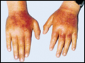 | Figure 358-4 Erythema and edema of the hands due to acute photosensitivity in a 10-year-old boy with erythropoietic protoporphyria. (From P. Poblette-Gutierrez et al.) |
view large |