

PART 16: Endocrinology and Metabolism
SECTION 2 Disorders of Bone and Mineral Metabolism
355 Paget's Disease and Other Dysplasias of Bone
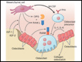 | Figure 355-1 Diagram illustrating factors that promote differentiation and function of osteoclasts and osteoblasts and the role of the RANK pathway. Stromal bone marrow (mesenchymal) cells and differentiated osteoblasts produce multiple growth factors and cytokines, including macrophage colony-stimulating factor (M-CSF), to modulate... |
view large |
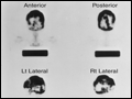 | Figure 355-2 A 48-year-old woman with Paget's disease of the skull. Left. Lateral radiograph showing areas of both bone resorption and sclerosis. Right.99mTc HDP bone scan with anterior, posterior, and lateral views of the skull showing diffuse isotope uptake by the frontal, parietal, occipital, and petrous... |
view large |
 | Figure 355-3 Radiograph of a 73-year-old man with Paget's disease of the right proximal femur. Note the coarsening of the trabecular pattern with marked cortical thickening and narrowing of the joint space consistent with osteoarthritis secondary to pagetic deformity of the right femur. |
view large |
 | Figure 355-4 Radiograph of a 16-year-old male with fibrous dysplasia of the right proximal femur. Note the multiple cystic lesions, including the large lucent lesion in the proximal midshaft with scalloping of the interior surface. The femoral neck contains two lucent cystic lesions. |
view large |