

PART 16: Endocrinology and Metabolism
SECTION 1 Endocrinology
339 Disorders of the Anterior Pituitary and Hypothalamus
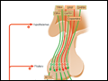 | Figure 339-1 Diagram of pituitary axes. Hypothalamic hormones regulate anterior pituitary trophic hormones that in turn determine target gland secretion. Peripheral hormones feed back to regulate hypothalamic and pituitary hormones. For abbreviations, see text. |
view large |
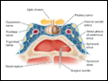
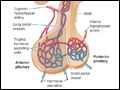 | Figure 339-2 Diagram of hypothalamic-pituitary vasculature. The hypothalamic nuclei produce hormones that traverse the portal system and impinge on anterior pituitary cells to regulate pituitary hormone secretion. Posterior pituitary hormones are derived from direct neural extensions. |
view large |
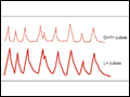 | Figure 339-3 Hypothalamic gonadotropin-releasing hormone (GnRH) pulses induce secretory pulses of luteinizing hormone (LH). |
view large |
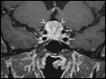 | Figure 339-4 Pituitary adenoma. Coronal T1-weighted postcontrast MR image shows a homogeneously enhancing mass (arrowheads) in the sella turcica and suprasellar region compatible with a pituitary adenoma; the small arrows outline the carotid arteries. |
view large |
 | Figure 339-5 Transsphenoidal resection of pituitary mass via the endonasal approach.(Adapted from R Fahlbusch: Endocrinol Metab Clin 21:669, 1992.) |
view large |
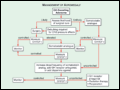
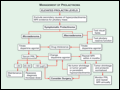 | Figure 339-6 Management of prolactinoma. MRI, magnetic resonance imaging; PRL, prolactin. |
view large |
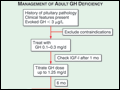 | Figure 339-7 Management of adult growth hormone (GH) deficiency. IGF, insulin-like growth factor. |
view large |
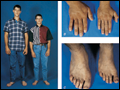 | Figure 339-8 Features of acromegaly/gigantism. A 22-year-old man with gigantism due to excess growth hormone is shown to the left of his identical twin. The increased height and prognathism (A) and enlarged hand (B) and foot |
view large |
 | Figure 339-9 Management of acromegaly. GH, growth hormone; CNS, central nervous system; IGF, insulin-like growth factor. (Adapted from S Melmed et al: J Clin Endocrinol Metab 94:1509–1517, 2009; © The Endocrine Society.) |
view large |
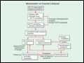 | Figure 339-10 Management of Cushing's syndrome. ACTH, adrenocorticotropin hormone; MRI, magnetic resonance imaging. *, Not usually required. |
view large |
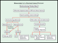 | Figure 339-11 Management of a nonfunctioning pituitary mass. |
view large |