

PART 15: Disorders of the Joints and Adjacent Tissues
SECTION 3 Disorders of the Joints and Adjacent Tissues
333 Gout and Other Crystal-Associated Arthropathies
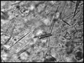 | Figure 333-1 Extracellular and intracellular monosodium urate crystals, as seen in a fresh preparation of synovial fluid, illustrate needle- and rod-shaped crystals. These crystals are strongly negative birefringent crystals under compensated polarized light microscopy; 400x. |
view large |
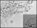 | Figure 333-2 Intracellular and extracellular calcium pyrophosphate dihydrate crystals, as seen in a fresh preparation of synovial fluid, illustrate rectangular, rod-shaped, and rhomboid crystals that are weakly positive birefringent crystals (compensated polarized light microscopy; 400x). |
view large |
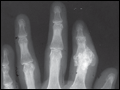 | Figure 333-3 A. Radiograph showing calcification due to apatite crystals surrounding an eroded joint. B. An electron micrograph demonstrates dark needle-shaped apatite crystals within a vacuole of a synovial fluid... |
view large |
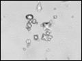 | Figure 333-4 Bipyramidal and small polymorphic calcium oxalate crystals from synovial fluid are a classic finding in CaOx arthropathy (ordinary light microscopy; 400x). |
view large |