

PART 15: Disorders of the Joints and Adjacent Tissues
SECTION 3 Disorders of the Joints and Adjacent Tissues
332 Osteoarthritis
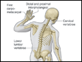 | Figure 332-1 Joints affected by osteoarthritis. |
view large |
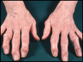 | Figure 332-2 Severe osteoarthritis of the hands affecting the distal interphalangeal joints (Heberden's nodes) and the proximal interphalangeal joints (Bouchard's nodes). There is no clear bony enlargement of the other common site in the hands, the thumb base. |
view large |
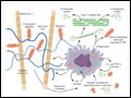 | Figure 332-3 The chondrocyte and its products, type II collagen, aggrecan, and enzymes, which degrade these structures along with molecules stimulating chondrocytes. IL, interleukin; NO, nitric oxide; OA, osteoarthritis; TNF, tumor necrosis factor. (From AR Poole et al: Ann Rheum... |
view large |
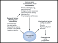 | Figure 332-4 Risk factors for osteoarthritis either contribute to the susceptibility of the joint (systemic factors or factors in the local joint environment) or increase risk by the load they put on the joint. Usually a combination of loading and susceptibility factors is required to cause disease or... |
view large |
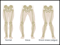 | Figure 332-5 The two types of limb malalignment in the frontal plane: varus, in which the stress is placed across the medial compartment of the knee joint, and valgus, which places excess stress across the lateral compartment of the knee. |
view large |
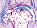 | Figure 332-6 Pathologic changes of osteoarthritis in a toe joint. Note the nonuniform loss of cartilage (arrowhead vs. solid arrow), the increased thickness of the subchondral bone envelope (solid arrow), and the... |
view large |
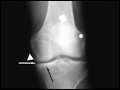 | Figure 332-7 X-ray of knee with medial osteoarthritis. Note the narrowed joint space on medial side of the joint only (white arrow), the sclerosis of the bone in the medial compartment providing evidence of cortical thickening (black arrow), and the... |
view large |