

PART 15: Disorders of the Joints and Adjacent Tissues
SECTION 3 Disorders of the Joints and Adjacent Tissues
331 Approach to Articular and Musculoskeletal Disorders
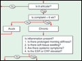 | Figure 331-1 Algorithm for the diagnosis of musculoskeletal complaints. An approach to formulating a differential diagnosis (shown in italics). CMC, carpometacarpal; CRP, C-reactive protein; DIP, distal interphalangeal; ESR, erythrocyte sedimentation rate; JA, juvenile arthritis; MCP,... |
view large |
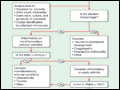
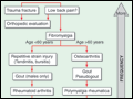 | Figure 331-2 Algorithm for consideration of the most common musculoskeletal conditions. GC, gonococcal; IBD, inflammatory bowel disease. |
view large |
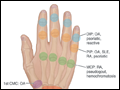 | Figure 331-3 Sites of hand or wrist involvement and their potential disease associations. CMC, carpometacarpal; DIP, distal interphalangeal; MCP, metacarpophalangeal; OA, osteoarthritis; PIP, proximal interphalangeal; RA, rheumatoid arthritis; SLE, systemic lupus erythematosus. |
view large |
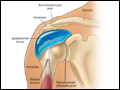 | Figure 331-4 Origins of shoulder pain. The schematic diagram of the shoulder indicates with arrows the most common causes and locations of shoulder pain. |
view large |
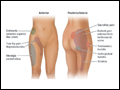 | Figure 331-5 Origins of hip pain and dysesthesias. (From Cush et al, with permission.) |
view large |
 | Figure 331-6 Algorithmic approach to the use and interpretation of synovial fluid aspiration and analysis. PMNs, polymorphonuclear (leukocytes); WBC, white blood cell (count). |
view large |
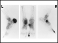 | Figure 331-7 [99mTc]Diphosphonate scintigraphy of the feet of a 33-year-old African-American male with reactive arthritis, manifested by sacroiliitis, urethritis, uveitis, asymmetric oligoarthritis, and enthesitis. This bone scan demonstrates increased uptake indicative of enthesitis involving... |
view large |
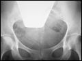 | Figure 331-8 Superior sensitivity of MRI in the diagnosis of osteonecrosis of the femoral head. A 45-year-old woman receiving highdose glucocorticoids developed right hip pain. Conventional x-rays (top) demonstrated only mild sclerosis of the right femoral head. T1-weighted... |
view large |