

PART 15: Disorders of the Joints and Adjacent Tissues
SECTION 2 Disorders of Immune-Mediated Injury
329 Sarcoidosis
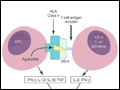 | Figure 329-1 Schematic representation of initial events of sarcoidosis. The antigen-presenting cell and helper T cell complex leads to the release of multiple cytokines. This forms a granuloma. Over time, the granuloma may resolve or lead to chronic disease, including fibrosis. APC, antigen-presenting... |
view large |
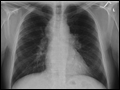 | Figure 329-2 Posterior-anterior chest roentgenogram demonstrating bilateral hilar adenopathy, stage 1 disease. |
view large |
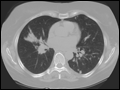 | Figure 329-3 High-resolution CT scan of chest demonstrating patchy reticular nodularity, including areas of confluence. |
view large |
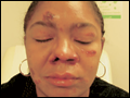 | Figure 329-4 Chronic inflammatory lesions around nose, eyes, and cheeks, referred to as lupus pernio. |
view large |
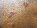 | Figure 329-5 Maculopapular lesions on the trunk of a sarcoidosis patient. |
view large |
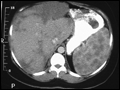 | Figure 329-6 CT scan of the abdomen after oral and intravenous contrast. The stomach is compressed by the enlarged spleen. Within the spleen, areas of hypo- and hyperdensity are identified. |
view large |
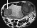 | Figure 329-7 MRI of wrist demonstrating large cyst in a sarcoidosis patient (line). |
view large |
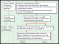 | Figure 329-8 Proposed approach to management of patient with possible sarcoidosis. Presence of one or more of these features supports the diagnosis of sarcoidosis: uveitis, optic neuritis, hypercalcemia, hypercalciuria, seventh cranial nerve paralysis, diabetes insipidus. |
view large |
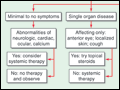 | Figure 329-9 The management of acute sarcoidosis is based on level of symptoms and extent of organ involvement. In patients with mild symptoms, no therapy may be needed unless specified manifestations are noted. |
view large |
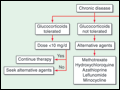 | Figure 329-10 Approach to chronic disease is based on whether glucocorticoid therapy is tolerated or not. |
view large |