

PART 15: Disorders of the Joints and Adjacent Tissues
SECTION 2 Disorders of Immune-Mediated Injury
323 Systemic Sclerosis (Scleroderma) and Related Disorders
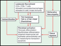 | Figure 323-1 Initial vascular injury in a genetically susceptible individual leads to functional and structural vascular alterations, inflammation, and autoimmunity. The inflammatory and immune responses initiate and sustain fibroblast activation and differentiation, resulting in pathologic... |
view large |
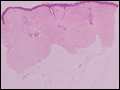 | Figure 323-2 Pathologic findings in systemic sclerosis (SSc). A. Dermal sclerosis. The skin is thickened due to marked expansion of the dermis. Thick bundles of densely packed collagen replace skin appendages. B. Early interstitial... |
view large |
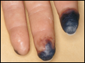 | Figure 323-3 Digital necrosis. Sharply demarcated necrosis of the fingertip in a patient with limited cutaneous systemic sclerosis (SSc) associated with severe Raynaud's phenomenon. |
view large |
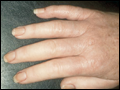 | Figure 323-4 Sclerodactyly. Note skin induration on the fingers, and fixed flexion contractures at the proximal interphalangeal joints in a patient with limited cutaneous systemic sclerosis (SSc). |
view large |
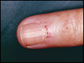 | Figure 323-5 Cutaneous vascular changes. A. Capillary changes at the nailfold in a patient with limited cutaneous systemic sclerosis (lcSSc). B. Telangiectasia on the face. |
view large |
 | Figure 323-6 Acro-osteolysis. Note dissolution of terminal phalanges in a patient with long-standing limited cutaneous systemic sclerosis (lcSSc) and Raynaud's phenomenon. |
view large |
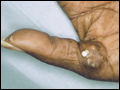 | Figure 323-7 Calcinosis cutis. Note large calcific deposit breaking through the skin in a patient with limited cutaneous systemic sclerosis (lcSSc). |
view large |
 | Figure 323-8 High-resolution CT scan of the lungs: interstitial lung disease. Note bilateral reticulonodular opacifications in a peripheral distribution in the lower lobes of the lungs in a patient with diffuse cutaneous systemic sclerosis (dcSSc). |
view large |