

PART 14: Disorders of the Gastrointestinal System
SECTION 3 Disorders of the Pancreas
313 Acute and Chronic Pancreatitis
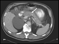 | Figure 313-1 Acute pancreatitis: CT evolution. A. Contrast-enhanced CT scan of the abdomen performed on admission for a patient with clinical and biochemical parameters suggestive of acute pancreatitis. Note the abnormal enhancement of the pancreatic parenchyma (arrow)... |
view large |
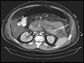 | Figure 313-2 A. Acute necrotizing pancreatitis: CT scan. Contrast-enhanced CT scan showing acute pancreatitis with necrosis. Arrow shows partially enhancing body/tail of pancreas surrounded by fluid with decreased enhancement in the neck/body of the pancreas. B. Acute fluid... |
view large |
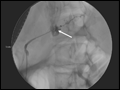 | Figure 313-3 A. Pancreaticopleural fistula: pancreatic duct leak on ERCP. Pancreatic duct leak demonstrated (arrow) at the time of retrograde pancreatogram in a patient with acute exacerbation of alcohol-induced acute or chronic pancreatitis. B.... |
view large |
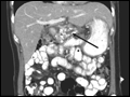 | Figure 313-4 A. Chronic pancreatitis and pancreatic calculi: CT scan. In this contrast-enhanced CT scan of the abdomen, there is evidence of an atrophic pancreas with multiple calcifications and stones in the parenchyma and dilated pancreatic duct (arrow). B. In this... |
view large |