

PART 14: Disorders of the Gastrointestinal System
SECTION 2 Liver and Biliary Tract Disease
304 Acute Viral Hepatitis
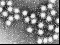 | Figure 304-1 Electron micrographs of hepatitis A virus particles and serum from a patient with hepatitis B. Left: 27-nm hepatitis A virus particles purified from stool of a patient with acute hepatitis A and aggregated by antibody to hepatitis A virus. ... |
view large |
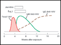 | Figure 304-2 Scheme of typical clinical and laboratory features of hepatitis A. |
view large |
 | Figure 304-3 Compact genomic structure of HBV. This structure, with overlapping genes, permits HBV to code for multiple proteins. The S gene codes for the “major” envelope protein, HBsAg. Pre-S1 and pre-S2, upstream of S, combine with S to code for two larger proteins, “middle”... |
view large |
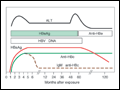
 | Figure 304-4 Scheme of typical clinical and laboratory features of acute hepatitis B. |
view large |
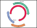
 | Figure 304-5 Scheme of typical laboratory features of wild-type chronic hepatitis B. HBeAg and HBV DNA can be detected in serum during the replicative phase of chronic infection, which is associated with infectivity and liver injury. Seroconversion from the replicative phase... |
view large |
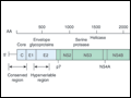 | Figure 304-6 Organization of the hepatitis C virus genome and its associated, 3000 amino-acid (AA) proteins. The three structural genes at the 5′ end are the core region, C, which codes for the nucleocapsid, and the envelope regions, E1 and E2, which code for envelope glycoproteins. The 5′... |
view large |
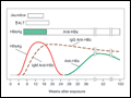
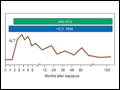 | Figure 304-7 Scheme of typical laboratory features during acute hepatitis C progressing to chronicity. HCV RNA is the first detectable event, preceding alanine aminotrans ferase (ALT) elevation and the appearance of anti-HCV. |
view large |