

PART 14: Disorders of the Gastrointestinal System
SECTION 1 Disorders of the Alimentary Tract
297 Diverticular Disease and Common Anorectal Disorders
 | Figure 297-1 Gross and microscopic view of sigmoid diverticular disease. Arrows mark an inflamed diverticulum with the diverticular wall made up only of mucosa. |
view large |
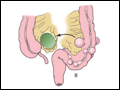 | Figure 297-2 Hinchey classification of diverticulitis. Stage I: Perforated diverticulitis with a confined paracolic abscess. Stage II: Perforated diverticulitis that has closed spontaneously with distant abscess formation. Stage III: Noncommunicating perforated diverticulitis with fecal peritonitis... |
view large |
 | Figure 297-3 Methods of surgical management of complicated diverticular disease. (1) Drainage, omental pedicle graft, and proximal diversion. (2) Hartmann's procedure. (3) Sigmoid resection with coloproctostomy. |
view large |
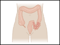
 | Figure 297-4 Degree of rectal prolapse. Mucosal prolapse only (A, B, sagittal view). Full-thickness prolapse associated with redundant rectosigmoid and deep pouch of Douglas (C, D, sagittal view). |
view large |
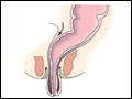
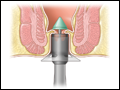 | Figure 297-5 Stapled transanal rectal resection. Schematic of placement of the circular stapling device. |
view large |
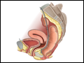 | Figure 297-6 Laparoscopic Ventral Rectopexy (LVR). To reduce the internal prolapse and close any rectovaginal septal defect, the pouch of Douglas is opened and mesh is secured to the anterolateral rectum, vaginal fornix, and sacrum. (From D'Hoore et al: Br J Surg 91:1500,... |
view large |
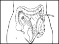
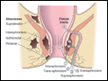 | Figure 297-7 Common locations of anorectal abscess (left) and fistula in ano (right). |
view large |