

PART 14: Disorders of the Gastrointestinal System
SECTION 1 Disorders of the Alimentary Tract
295 Inflammatory Bowel Disease
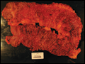 | Figure 295-1 Ulcerative colitis. Diffuse (nonsegmental) mucosal disease, with broad areas of ulceration. The bowel wall is not thickened, and there is no cobblestoning. (Courtesy of Dr. R Odze, Division of Gastrointestinal Pathology, Department of Pathology, Brigham and Women's... |
view large |
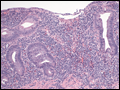 | Figure 295-2 Medium power view of colonic mucosa in ulcerative colitis showing diffuse mixed inflammation, basal lymphoplasmacytosis, crypt atrophy and irregularity and superficial erosion. These features are typical of chronic active ulcerative colitis. (Courtesy of Dr. R Odze,... |
view large |
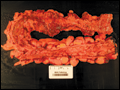 | Figure 295-3 Crohn's disease of the colon showing thickening of the wall, with stenosis, linear serpiginous ulcers and cobblestoning of the mucosa. (Courtesy of Dr. R Odze, Division of Gastrointestinal Pathology, Department of Pathology, Brigham and Women's Hospital, Boston,... |
view large |
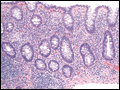 | Figure 295-4 Medium power view of Crohn's colitis showing mixed acute and chronic inflammation, crypt atrophy, and multiple small epithelioid granulomas in the mucosa. (Courtesy of Dr. R Odze, Division of Gastrointestinal Pathology, Department of Pathology, Brigham and Women's... |
view large |
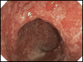 | Figure 295-5 Colonoscopy with acute ulcerative colitis: Severe colon inflammation with erythema, friability, and exudates. (Courtesy of Dr. M. Hamilton, Gastroenterology Division, Department of Medicine, Brigham and Women's Hospital, Boston, Massachusetts; with permission.) |
view large |
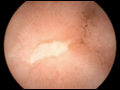 | Figure 295-6 Wireless capsule endoscopy image in a patient with Crohn's disease of the ileum shows ulcerations and narrowing of the intestinal lumen. (Courtesy of Dr. S Reddy, Gastroenterology Division, Department of Medicine, Brigham and Women's Hospital, Boston, Massachusetts;... |
view large |
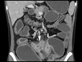 | Figure 295-7 Coronal contrast-enhanced multidetector computed tomography (MDCT) image obtained after oral administration of 1350 cc of neutral oral contrast material shows dilation of small bowel loops, segmental mucosal hyperenhancement, and interloop sinus tracts (white... |
view large |
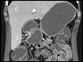 | Figure 295-8 Coronal contrast-enhanced multidetector computed tomography (MDCT) image obtained after oral administration of 1350 cc of neutral oral contrast material shows mucosal hyperenhancement of the terminal ileum with narrowing and mild prestenotic dilatation. (Courtesy of... |
view large |
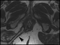 | Figure 295-9 Axial T2-weighted MR image obtained in a 37-year-old man with Crohn's disease shows a linear fluid-filled perianal fistula (arrow) in the right ischioanal fossa. (Courtesy of Dr. K Mortele, Gastrointestinal Radiology, Department of... |
view large |
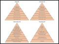 | Figure 295-10 Medical management of IBD. 5-ASA, 5-aminosalicylic acid; CD, Crohn's disease; UC, ulcerative colitis. |
view large |
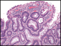 | Figure 295-11 Medium power view of low-grade dysplasia in a patient with chronic ulcerative colitis. Low-grade dysplastic crypts are interspersed among regenerating crypts. (Courtesy of Dr. R Odze, Division of Gastrointestinal Pathology, Department of Pathology, Brigham and Women's... |
view large |