

PART 14: Disorders of the Gastrointestinal System
SECTION 1 Disorders of the Alimentary Tract
292 Diseases of the Esophagus
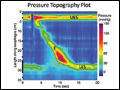 | Figure 292-1 High-resolution esophageal pressure topography (right) and conventional manometry (left) of a normal swallow. LES, lower esophageal sphincter; E, esophageal body; UES, upper esophageal sphincter. |
view large |
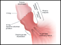 | Figure 292-2 Radiographic anatomy of the gastroesophageal junction. |
view large |
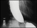
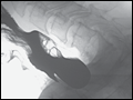 | Figure 292-3 Examples of small (left) and large (middle, right) Zenker's diverticulum arising from Killian's triangle in the distal hypopharynx. Smaller diverticula are evident only during the swallow, whereas larger ones retain food and fluid. |
view large |
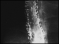 | Figure 292-4 Intramural esophageal pseudodiverticulosis associated with chronic obstruction. Invaginations of contrast into the esophageal wall outline deep esophageal glands. |
view large |
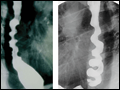
 | Figure 292-5 Achalasia with esophageal dilatation, tapering at the gastroesophageal junction and an air-fluid level within the esophagus. The example on the left shows sigmoid deformity with very advanced disease. |
view large |
 | Figure 292-6 Three subtypes of achalasia: classic (Panel A), with esophageal compression (Panel B), and spastic achalasia (Panel C) imaged with pressure topography. All are characterized by impaired lower esophageal sphincter (LES) relaxation and absent... |
view large |
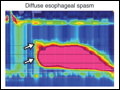
 | Figure 292-7 Diffuse esophageal spasm. The characteristic “corkscrew” esophagus results from spastic contraction of the circular muscle in the esophageal wall; more precisely, this is actually a helical array of muscle. These findings are also seen with spastic achalasia. |
view large |
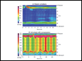
 | Figure 292-8 Esophageal pressure topography of the two major variants of esophageal spasm: spastic nutcracker (left) and diffuse esophageal spasm (right). Spastic nutcracker is defined by the extraordinarily vigorous and repetitive contractions with normal peristaltic onset. Diffuse... |
view large |
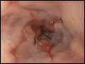 | Figure 292-9 Endoscopic appearance of (A) peptic esophagitis, (B) a peptic stricture, (C) Barrett's metaplasia, and (D) adenocarcinoma developing within an area of Barrett's esophagus. |
view large |
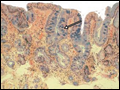 | Figure 292-10 Histopathology of Barrett's metaplasia and Barrett's with high-grade dysplasia. H&E, hematoxylin and eosin. |
view large |
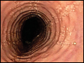 | Figure 292-11 Endoscopic features of (A) eosinophilic esophagitis (EoE), (B) Candida esophagitis, (C) giant ulcer associated with HIV, (D) and a Schatzki ring. |
view large |
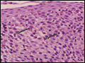 | Figure 292-12 Histopathology of eosinophilic esophagitis (EoE) showing dense infiltration of the esophageal squamous epithelium with eosinophils. Eosinophilic inflammation can also be seen with gastroesophageal reflux disease (GERD); the optimal discriminatory threshold for EoE is greater than 15 eosinophils per high-power field. |
view large |