

PART 13: Disorders of the Kidney and Urinary Tract
284 Polycystic Kidney Disease and Other Inherited Tubular Disorders
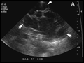 | Figure 284-1 Renal ultrasonogram and contrast-enhanced abdominal CT scan in a 56-year-old woman with autosomal dominant polycystic kidney disease. A. Sonogram of the right kidney showing numerous cysts of varying sizes (arrows). B. Abdominal CT scan... |
view large |
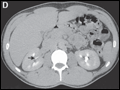 | Figure 284-2 Radiographs of medullary sponge kidney disease. A. Plain x-ray film of a patient with a history of recurrent nephrolithiasis showing clusters of stones in the papillae (arrows). B–E. CT scan of an 18-year-old male patient investigated for persistent microscopic... |
view large |
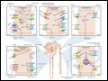 | Figure 284-3 Schematic representation of channels, transporters, and enzymes associated with hereditary renal tubular disorders. AD, autosomal dominant; AR, autosomal recessive; DI, diabetes insipidus; NKCC2, Na-K-2Cl co-transporter; ROMK, renal outer medullary potassium channel; AQP2, aquaporin-2; CLC-Kb, chloride channel Kb; CaR,... |
view large |