

PART 13: Disorders of the Kidney and Urinary Tract
280 Chronic Kidney Disease
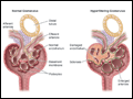 | Figure 280-1 Left: Schema of the normal glomerular architecture. Right: Secondary glomerular changes associated with a reduction in nephron number, including enlargement of capillary lumens and focal adhesions, which are thought to occur consequent to compensatory hyperfiltration and hypertrophy in the... |
view large |
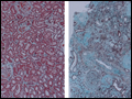 | Figure 280-2 Left: Low-power photomicrograph of a normal kidney showing normal glomeruli and healthy tubulointerstitium without fibrosis. Right: Low-power photomicrograph of chronic kidney disease with sclerosis of many glomeruli and severe tubulointerstitial fibrosis (Masson... |
view large |
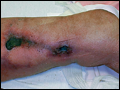 | Figure 280-3 Calciphylaxis. This peritoneal dialysis patient was on chronic warfarin therapy for prophylactic anticoagulation for a mechanical heart valve. She slept with the dialysis catheter pressed between her legs. A small abrasion was followed by progressive skin necrosis along the catheter tract... |
view large |
 | Figure 280-4 U.S. Renal Data System showing increased likelihood of dying rather than starting dialysis or reaching stage 5 chronic kidney disease (CKD). 1, Death; 2, ESRD/D; 3, event-free. DM; diabetes mellitus. (Adapted from RN Foley et al: J Am Soc Nephrol 16:489-95, 2005.) |
view large |