

PART 12: Critical Care Medicine
SECTION 4 Oncologic Emergencies
275 Neurologic Critical Care, Including Hypoxic-Ischemic Encephalopathy, and Subarachnoid Hemorrhage
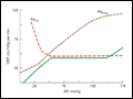 | Figure 275-1 Autoregulation of cerebral blood flow (solid line). Cerebral perfusion is constant over a wide range of systemic blood pressure. Perfusion is increased in the setting of hypoxia or hypercarbia. BP, blood pressure; CBF, cerebral blood flow.... |
view large |
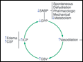 | Figure 275-2 Ischemia and vasodilatation. Reduced cerebral perfusion pressure (CPP) leads to increased ischemia, vasodilation, increased intracranial pressure (ICP), and further reductions in CPP, a cycle leading to further neurologic injury. CBV, cerebral blood volume; CMR, cerebral... |
view large |
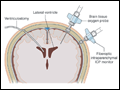 | Figure 275-3 Intracranial pressure and brain tissue oxygen monitoring. A ventriculostomy allows for drainage of cerebrospinal fluid to treat elevated intracranial pressure (ICP). Fiberoptic ICP and brain tissue oxygen monitors are usually secured using a screwlike skull bolt. Cerebral blood... |
view large |
 | Figure 275-4 Prognostication of outcome in comatose survivors of cardiopulmonary resuscitation. Numbers in parentheses are 95% confidence intervals. Confounders could include use of sedatives or neuromuscular blocking agents, hypothermia therapy, organ failure, or shock. Tests denoted with an... |
view large |
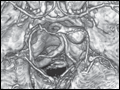
 | Figure 275-5 Cortical laminar necrosis in hypoxic-ischemic encephalopathy. T1-weighted postcontrast MRI shows cortical enhancement in a watershed distribution consistent with laminar necrosis. |
view large |
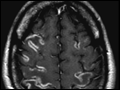
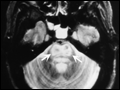 | Figure 275-6 Central pontine myelinolysis. Axial T2-weighted MR scan through the pons reveals a symmetric area of abnormal high signal intensity within the basis pontis (arrows). |
view large |
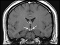 | Figure 275-7 Wernicke's disease. Coronal T1-weighted postcontrast MRI reveals abnormal enhancement of the mammillary bodies (arrows), typical of acute Wernicke's encephalopathy. |
view large |
 | Figure 275-8 Subarachnoid hemorrhage. A. CT angiography revealing an aneurysm of the left superior cerebellar artery. B. Noncontrast CT scan at the level of the third ventricle revealing... |
view large |