

PART 12: Critical Care Medicine
SECTION 1 Respiratory Critical Care
267 Approach to the Patient With Critical Illness
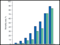 | Figure 267-1 APACHE II survival curve. Blue, nonoperative; green, postoperative. |
view large |
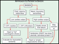 | Figure 267-2 Approach to patient in shock. EGDT, early goal-directed therapy; JVP, jugular venous pulse. |
view large |
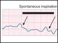 | Figure 267-3 Right atrial pressure change during spontaneous respiration in a patient with shock who will increase cardiac output in response to intravenous fluid administration. The right atrial pressure decreases from 7 mmHg to 4 mmHg. The horizontal bar marks the time of spontaneous... |
view large |
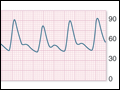 | Figure 267-4 Pulse pressure change during mechanical ventilation in a patient with shock who will increase cardiac output in response to intravenous fluid administration. The pulse pressure (systolic minus diastolic blood pressure) changes during mechanical ventilation in a patient with... |
view large |
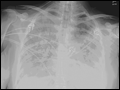 | Figure 267-5 Chest radiograph of a patient with ARDS. ARDS, acute respiratory distress syndrome. |
view large |
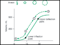 | Figure 267-6 Pressure-volume relationship of the lungs of a patient with ARDS. At the lower inflection point, collapsed alveoli begin to open, and the lung compliance changes. At the upper deflection point, alveoli become overdistended. The shape and size of alveoli are illustrated at the... |
view large |
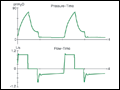 | Figure 267-7 Increased airway resistance with autoPEEP. The top waveform (airway pressure vs. time) shows a large difference between the peak airway pressure (80 cmH2O) and the plateau airway pressure (20 cmH2O). The bottom waveform (flow vs. time) demonstrates airflow... |
view large |