

PART 11: Disorders of the Respiratory System
SECTION 2 Diseases of the Respiratory System
258 Bronchiectasis and Lung Abscess
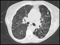 | Figure 258-1 Representative chest CT image of severe bronchiectasis. This patient's CT demonstrates many severely dilated airways, seen both longitudinally (arrowhead) and in cross-section (arrow). |
view large |
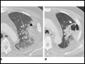 | Figure 258-2 Representative chest CT demonstrating development of lung abscesses. This patient was immunocompromised due to underlying lymphoma and developed severe Pseudomonas aeruginosa pneumonia, as represented by a left lung infiltrate with concern for central regions of... |
view large |