

PART 11: Disorders of the Respiratory System
SECTION 2 Diseases of the Respiratory System
256 Occupational and Environmental Lung Disease
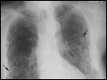 | Figure 256-1 Asbestosis: A. Frontal chest radiograph shows bilateral calcified pleural plaques consistent with asbestos-related pleural disease. Poorly defined linear and reticular abnormalities are seen in the lower lobes bilaterally. |
view large |
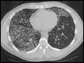 | Figure 256-2 Acute silicosis. This high-resolution computed tomography scan shows multiple small nodules consistent with silicosis but also diffuse ground-glass densities with thickened intralobular and interlobular septa producing polygonal shapes. This has been referred to as "crazy paving." |
view large |
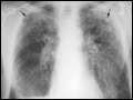 | Figure 256-3 Chronic silicosis. A. Frontal chest radiograph in a patient with silicosis shows variably sized, poorly defined nodules (arrows) predominating in the upper lobes, |
view large |
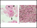 | Figure 256-4 Histopathologic features of biomass smoke–induced interstitial lung disease.A. Anthracitic pigment is seen accumulating along alveolar septae (arrowheads) and within a pigmented dust macule ( |
view large |