

PART 11: Disorders of the Respiratory System
SECTION 2 Diseases of the Respiratory System
255 Hypersensitivity Pneumonitis and Pulmonary Infiltrates with Eosinophilia
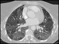 | Figure 255-1 Chest CT scan of a patient with subacute hypersensitivity pneumonitis in which scattered regions of ground-glass infiltrates in a mosaic pattern consistent with air trapping are seen bilaterally. This patient had bird fancier's lung.... |
view large |
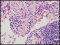 | Figure 255-2 Open-lung biopsy from a patient with subacute hypersensitivity pneumonitis demonstrating a loose, nonnecrotizing granuloma made up of histiocytes and multinucleated giant cells. Peribronchial inflammatory infiltrate made up of lymphocytes and plasma cells is also seen. |
view large |