

PART 11: Disorders of the Respiratory System
SECTION 1 Diagnosis of Respiratory Disorders
253 Diagnostic Procedures in Respiratory Disease
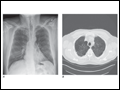 | Figure 253-1 Chest x-ray (A) and CT scan (B) from a patient with emphysema. The extent and distribution of emphysema are not well appreciated on plain film but clearly evident on CT scan obtained. |
view large |
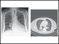 | Figure 253-2 Chest x-ray (A) and CT scan (B) demonstrating a right lower-lobe mass. The mass is not well appreciated on the plain film because of the hilar structures and known calcified adenopathy. CT is superior to plain radiography for the detection of abnormal mediastinal densities and... |
view large |
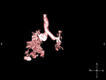 | Figure 253-3 Spiral CT with reconstruction of images in planes other than axial view. Spiral CT in a lung transplant patient with a dehiscence and subsequent aneurysm of the anastomosis. CT images were reconstructed in the sagittal view (A) and using... |
view large |
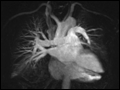
 | Figure 253-4 MRA image of the vasculature of a patient after lung transplant. The image demonstrates the detailed view of the vasculature that can be obtained using digital subtraction techniques. Images from a patient after lung transplant show the venous and arterial anastomosis on the... |
view large |
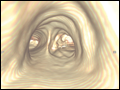
 | Figure 253-5 Virtual bronchoscopic image of the trachea. The view projected is one that would be obtained from the trachea looking down to the carina. The left and right main stem airways are seen bifurcating from the carina. |
view large |