

PART 10: Disorders of the Cardiovascular System
SECTION 4 Disorders of the Heart
239 Pericardial Disease
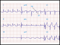 | Figure 239-1 Acute pericarditis often produces diffuse ST-segment elevations (in this case in leads I, II, aVF, and V2 to V6) due to a ventricular current of injury. Note also the characteristic PR-segment deviation (opposite in polarity to the ST segment) due to a concomitant atrial i ... |
view large |
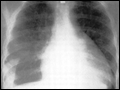 | Figure 239-2 Chest radiogram from a patient with a pericardial effusion showing typical “water bottle” heart. There is also a right pleural effusion. [From SS Kabbani, M LeWinter, in MH Crawford et al (eds): Cardiology. London, Mosby, 2001.] |
view large |
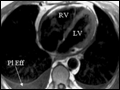
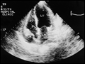 | Figure 239-3 Apical four-chamber echocardiogram recorded in a patient with a moderate pericardial effusion and evidence of hemodynamic compromise. The frame is recorded in early ventricular systole, immediately after atrial contraction. Note that the right atrial wall is indented inward and its curvatur ... |
view large |
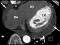 | Figure 239-4 Chronic pericardial effusion in a 54-year-old female patient with Hodgkin's disease seen in contrast-enhanced 64-slice CT. The arrows point at the pericardial effusion (LV, left ventricle; RV, right ventricle; RA, right atrium). Due to the timing of the scan relative to contrast injection, ... |
view large |
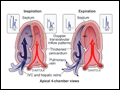 | Figure 239-5 Constrictive pericarditis. Doppler schema of respirophasic changes in mitral and tricuspid inflow. Reciprocal patterns of ventricular filling are assessed on pulsed Doppler examination of mitral valve (MV) and tricuspid valve (TV) inflow. |
view large |
 | Figure 239-6 Cardiovascular magnetic resonance in a patient with constrictive pericarditis. On the right is a basal short-axis view of the ventricles showing a thickened pericardium encasing the heart (arrows). On the left is a transaxial view, again showing the thickened peri ... |
view large |