

PART 10: Disorders of the Cardiovascular System
SECTION 4 Disorders of the Heart
236 Congenital Heart Disease in the Adult
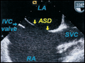 | Figure 236-1 Secundum atrial septal defect. Transesophageal echocardiogram of secundum ASD and device closure. A. The atrial septal defect (ASD) between the left atrium (LA) and right atrium (RA) is shown. B. A percutaneous catheter–deliver ... |
view large |
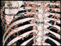 | Figure 236-2 Aortic coarctation. The extensive collaterals (left) underneath the ribs and in the periscapular region are shown on a posterior view of a three-dimensional CT angiogram, which are responsible for rib notching on chest x-ray. dao, descending aorta. |
view large |
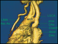 | Figure 236-3 Aortic coarctation. The coarctation (Coarct) of the aorta is shown in the typical “adult” location in the descending aorta (DAo) just distal to the dilated left subclavian artery (LSCA) in this three-dimensional reconstruction of an MR angiogram. There is a post-coarct aneurysm ... |
view large |
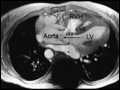 | Figure 236-4 Tetralogy of Fallot. Magnetic resonance angiogram. A mid-systolic frame showing the malaligned ventricular septal defect (VSD) with the aorta overriding the ventricular septal defect. LV, left ventricle; RVH, RV hypertrophy; VS, ventricular septum. |
view large |