

PART 10: Disorders of the Cardiovascular System
SECTION 3 Disorders of Rhythm
233 The Tachyarrhythmias
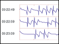 | Figure 233-1 Spontaneous termination of atrial fibrillation at the time of a syncopal episode identified from implantable loop ECG recording. |
view large |
 | Figure 233-2 Schematic representation of the different mechanisms for arrhythmias. A. Abnormal automaticity due to an increased slope of phase 4 of the action potential or a decrease in the threshold for phase 0. B. Triggered activity due to early... |
view large |
 | Figure 233-3 Atrial and ventricular premature complexes (APCs, VPCs). The APC resets the sinus node, and no compensatory pause is present A. even when conducted aberrantly in the ventricles with a bundle branch... |
view large |
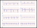 | Figure 233-4 Supraventricular tachycardias with irregular ventricular rates. Atrial fibrillation A. atrial flutter B. atrial tachycardia... |
view large |
 | Figure 233-5 Atrial fibrillation A. transitions to "slow" atrial flutter during antiarrhythmic drug therapy. B. A rapid ventricular... |
view large |
 | Figure 233-6 Atrial flutter/atrial fibrillation. Coarse atrial fibrillation A. contrasted with organized atrial flutter B. |
view large |
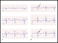
 | Figure 233-7 Pattern of atrial and ventricular activation and characteristic relationship of P-wave and QRS complex as recorded in leads II and V1 during regular supraventricular tachycardias. A. Sinus... |
view large |
 | Figure 233-8 A. Sinus rhythm tracing of leads V1–V3 showing evidence of Wolff-Parkinson-White syndrome with short PR interval and delta wave. B. During... |
view large |
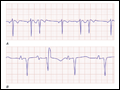
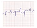
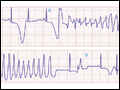 | Figure 233-9 Sinus rhythm with long QT interval and the polymorphic ventricular arrhythmia torsades des pointes. Dramatic T wave alternans is present in sinus rhythm. |
view large |
 | Figure 233-10 Ventricular tachycardia. ECG showing AV dissociation (arrows mark P waves), wide QRS >200 ms, superior frontal plane axis, slurring of the initial portion of the QRS, and large S wave in V6—all clues to the diagnosis of ventricular tachycardia. |
view large |
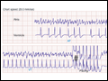 | Figure 233-11 Ventricular tachycardia (VT) (*) during atrial fibrillation stopped by pacing (#) from an implantable cardioverter defibrillator (ICD) from recording stored by ICD. The atrial electrogram shows characteristic fibrillatory waves through the tracing. The ventricular electrogram... |
view large |
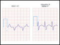 | Figure 233-12 Common idiopathic ventricular tachycardia (VT) ECG patterns. Right ventricular outflow tract (RVOT) VT with typical left bundle QRS pattern in V1 and inferiorly directed frontal plane axis, and left ventricular septal VT from the inferior septum with a narrow QRS RBBB... |
view large |
 | Figure 233-13 Bundle branch reentrant ventricular tachycardia (VT) showing typical QRS morphologies when VT is initiated with stimulation from the right ventricle [left bundle branch block (LBBB) VT pattern] or left ventricle [right bundle branch block (RBBB) VT pattern] and schema for circuit... |
view large |
 | Figure 233-14 Leads V1 to V3 in sinus rhythm from a normal subject A., from a patient with arrhythmogenic right ventricular cardiomyopathy showing epsilon waves (arrow) and T-wave inversion ... |
view large |
 | Figure 233-15 Digoxin toxic bidirectional fascicular tachycardia. |
view large |
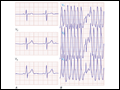
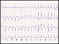
 | Figure 233-16 Catecholaminergic polymorphic ventricular tachycardia noted during an exercise stress test. |
view large |