

PART 10: Disorders of the Cardiovascular System
SECTION 3 Disorders of Rhythm
231 Principles of Electrophysiology
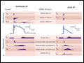 | Figure 231-1 A. Cellular atrial and ventricular action potentials. Phases 0–4 are the rapid upstroke, early repolarization, plateau, late repolarization, and diastole, respectively. The ionic currents and their respective genes are shown above and below the action poten ... |
view large |
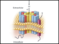 | Figure 231-2 Topology and subunit composition of the voltage-dependent ion channels. Potassium channels are formed by the tetramerization of α or pore-forming subunits and one or more β subunits; only single β subunits are shown for clarity. Sodium and calcium channels are comp ... |
view large |
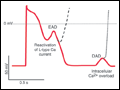 | Figure 231-3 Schematic action potentials with early after depolarizations (EADs) and delayed afterdepolarizations (DADs). Afterdepolarizations are spontaneous depolarizations in cardiac myocytes. EADs occur before the end of the action potential (phases 2 and 3), interrupting repolarization. D ... |
view large |
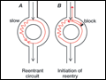 | Figure 231-4 Schematic diagram of reentry. A. The circuit contains two limbs, one with slow conduction. B. A premature impulse blocks in the fast pathway and conducts over the slow pathway, allowing ... |
view large |
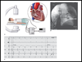 | Figure 231-5 Catheter ablation of cardiac arrhythmias. A. A schematic of the catheter system and generator in a patient undergoing radiofrequency catheter ablation (RFCA); the circuit involves the catheter in the heart and a dispersive pa ... |
view large |