

PART 10: Disorders of the Cardiovascular System
SECTION 2 Diagnosis of Cardiovascular Disorders
230 Diagnostic Cardiac Catheterization and Coronary Angiography
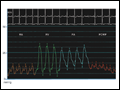 | Figure 230-1 Normal hemodynamic waveforms recorded during right heart catheterization. Atrial pressure tracings have a characteristic "a" wave that reflects atrial contraction and a "v" wave that reflects pressure changes in the atrium during ventricular ... |
view large |
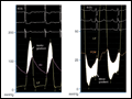 | Figure 230-2 Severe aortic and mitral stenosis. Simultaneous recording of left ventricular (LV) and aortic (Ao) pressure tracings demonstrate a 62-mmHg mean systolic gradient (shaded area) that corresponds to an aortic valve area of 0.6 cm2 (left). Simultaneous recording of LV and pul ... |
view large |
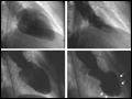 | Figure 230-3 Left ventriculogram at end diastole (left) and end systole (right). In patients with normal left ventricular function, the ventriculogram reveals symmetric contraction of all walls (top). Patients with coronary artery disease may have wall motion abnormalities on ventriculography ... |
view large |
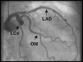 | Figure 230-4 Normal coronary artery anatomy. A. Coronary angiogram showing the left circumflex (LCx) artery and its obtuse marginal (OM) branches. The left anterior descending artery (LAD) is also seen but may be fore ... |
view large |
 | Figure 230-5 Coronary stenoses on cine angiogram and intravascular ultrasound. Significant stenoses in the coronary artery are seen as narrowings (black arrows) of the vessel. Intravascular ultrasound shows a normal segment of artery (A), areas with eccentric plaque (B, C), and near total obli ... |
view large |