

PART 8: Infectious Diseases
SECTION 18 Protozoal Infections
209 Amebiasis and Infection With Free-Living Amebas
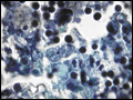
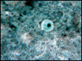 | Figure 209-1 Entamoeba cyst. Three of the four nuclei are clearly visible. (Courtesy of Dr. George Healy, Centers for Disease Control and Prevention.) |
view large |
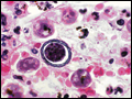
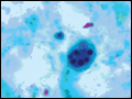 | Figure 209-2 E. histolytica trophozoite with ingested red blood cells. Note the single nucleus with central nucleolus. (Courtesy of the Centers for Disease Control and Prevention.) |
view large |
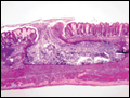 | Figure 209-3 E. histolytica flask-shaped intestinal ulceration from a kitten. (Courtesy of Dr. Mae Melvin, Centers for Disease Control and Prevention.) |
view large |
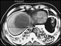 | Figure 209-4 Large amebic abscess in the right lobe of the liver visualized by CT. (Courtesy of Dr. M. M. Reeder, International Registry of Tropical Imaging.) |
view large |
 | Figure 209-5 Naegleria in a section of human brain tissue from a patient with primary amebic meningoencephalitis. (Courtesy of Dr. George Healy, Centers for Disease Control and Prevention.) |
view large |
 | Figure 209-6 Acanthamoeba cyst in brain tissue from a patient with granulomatous amebic encephalitis. (Courtesy of Dr. George Healy, Centers for Disease Control and Prevention.) |
view large |