

PART 8: Infectious Diseases
SECTION 15 Infections Due to RNA Viruses
194 Mumps
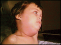 | Figure 194-1 Child with mumps. Note the classic submandibular and preauricular enlargement of the parotid gland. (From the Centers for Disease Control and Prevention.) |
view large |
 | Figure 194-2 Schematic drawing of a parotid gland infected with mumps virus (right) compared with a normal gland (left). An enlarged cervical lymph node is usually posterior to the imaginary line. (Reprinted with permission from Gershon A et al: Mumps, in Krugman's Infectious Diseases of Children, 11th ed. Philadelphia, Elsevier, 2004, p... |
view large |