

PART 8: Infectious Diseases
SECTION 15 Infections Due to RNA Viruses
189 Human Immunodeficiency Virus Disease: AIDS and Related Disorders
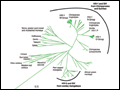 | Figure 189-1 A phylogenetic tree based on the complete genomes of primate immunodeficiency viruses. The scale (0.10) indicates a 10% difference at the nucleotide level. (Prepared by Brian Foley, PhD, of the HIV Sequence Database, Theoretical Biology and Biophysics Group, Los Alamos National Laboratory; additional... |
view large |
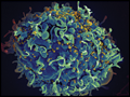 | Figure 189-2 A. Electron micrograph of HIV. Figure illustrates a typical virion following budding from the surface of a CD4+ T lymphocyte, together with two additional incomplete virions in the process of budding from the cell membrane. ... |
view large |
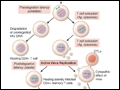
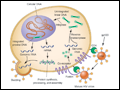 | Figure 189-3 The replication cycle of HIV. See text for description. (Adapted from AS Fauci: Nature 384:529, 1996.) |
view large |
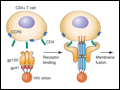 | Figure 189-4 Binding and fusion of HIV-1 with its target cell. HIV-1 binds to its target cell via the CD4 molecule, leading to a conformational change in the gp120 molecule that allows it to bind to the co-receptor CCR5 (for R5-using viruses). The virus then firmly attaches to the host cell membrane in a coiled-spring fashion via the newly... |
view large |
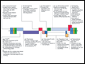 | Figure 189-5 Organization of the genome of the HIV provirus together with a summary description of its 9 genes encoding 15 proteins. (Adapted from Greene and Peterlin.) |
view large |
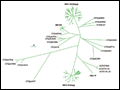 | Figure 189-6 Phylogenetic tree constructed from representative viral envelope sequences of the subtypes and CRF01 in HIV-1 group M, some isolates from groups N, O, and P (also human HIV-1), CPZ (chimpanzee), and gorilla (GOR). The scale bar indicates the genetic distances between the sequences. (Prepared by Brian Foley,... |
view large |
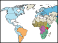 | Figure 189-7 Geographic distribution of HIV-1 subtypes and recombinant forms. (Adapted from Taylor et al; with permission.) |
view large |
 | Figure 189-8 Probability of HIV transmission per coital act among monogamous, heterosexual, HIV-serodiscordant couples in Uganda. (From RH Gray et al.) |
view large |
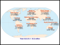 | Figure 189-9 Estimated number of adults and children living with HIV infection as of December, 2009. Total: 33.3 (31.4–35.3) (31.1–35.8) million. [From Joint United Nations Programme on HIV/AIDS (UNAIDS).] |
view large |
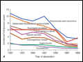
 | Figure 189-10 Global HIV/AIDS epidemiologic estimates, 1990–2009. A. Number of people living with HIV. B. Number of people newly infected with HIV globally. C. Number of adult and child deaths due to AIDS. (From... |
view large |
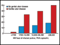
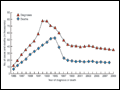 | Figure 189-11 Estimated number of AIDS cases and AIDS deaths, United States, 1985–2009. (From CDC.) |
view large |
 | Figure 189-13 Transmission categories of adults and adolescents with HIV/AIDS diagnosed during 2009 in the United States. Estimates from 40 states with confidential, name-based HIV infection reporting. Data include persons with a diagnosis of HIV infection regardless of AIDS status at diagnosis. (From CDC.) |
view large |
 | Figure 189-14 Estimated AIDS diagnoses among adults and adolescents, by transmission category and year of diagnosis, United States, 1985–2009. (From CDC.) |
view large |
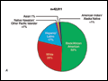 | Figure 189-15 Race/ethnicity of persons (including children) with HIV/AIDS diagnosed during 2009 in the United States. A. Proportion of new infections by race/ethnicity. B. Rate of new infections by race/ethnicity (per 100,000... |
view large |
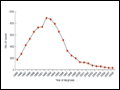 | Figure 189-16 Estimated numbers of perinatally acquired AIDS cases in children by year of diagnosis, 1985–2009, United States (From CDC.) |
view large |
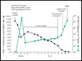 | Figure 189-17 Typical course of an untreated HIV-infected individual. See text for detailed description. (From G Pantaleo et al: N Engl J Med 328:327, 1993. Copyright 1993 Massachusetts Medical Society. All rights reserved.) |
view large |
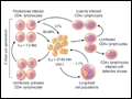
 | Figure 189-18 Summary of early events in HIV infection. See text for detailed description. CTLs, cytolytic T lymphocytes; HIV, human immunodeficiency virus. (Adapted from Haase, 2005.) |
view large |
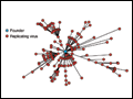 | Figure 189-19 As HIV diverges from founder to chronically replicating virus, it accumulates N-linked glycosylation sites. See text for detailed description. (Adapted from CA Derdeyn et al: Science 303:2019, 2004; B Chohan et al:J Virol 79:6528, 2005; and BF Keele et al:Proc Natl Acad Sci USA105:7552,... |
view large |
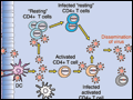
 | Figure 189-20 Generation of latently infected, resting CD4+ T cells in HIV-infected individuals. See text for details. Ag, antigen; CTLs, cytolytic T lymphocytes. (Courtesy of TW Chun; with permission.) |
view large |
 | Figure 189-21 Dynamics of HIV infection in vivo. See text for detailed description. (From AS Perelson et al:Science271:1582, 1996.) |
view large |
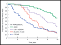 | Figure 189-22 Relationship between levels of virus and rates of disease progression. Kaplan-Meier curves for AIDS-free survival stratified by baseline HIV-1 RNA categories (copies per milliliter). (From Mellors et al.) |
view large |
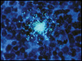 | Figure 189-23 HIV in the lymph node of an HIV-infected individual. An individual cell infected with HIV shown expressing HIV RNA by in situ hybridization using a radiolabeled molecular probe. Original ×500. (Adapted from G Pantaleo, AS Fauci.) |
view large |
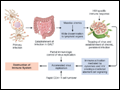 | Figure 189-24 Events that transpire from primary HIV infection through the establishment of chronic persistent infection to the ultimate destruction of the immune system. See text for details. CTLs, cytolytic T lymphocytes; GALT, gut-associated lymphoid tissue. |
view large |
 | Figure 189-25 Model for the role of co-receptors CXCR4 and CCR5 in the efficient binding and entry of X4 (A) and R5 (B) strains of HIV-1, respectively, into CD4+ target cells. Blocking of this initial event in the virus life cycle can... |
view large |
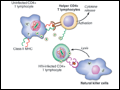 | Figure 189-26 Schematic representation of the different immunologic effector mechanisms thought to be active in the setting of HIV infection. Detailed descriptions are given in the text. ADCC, antibody-dependent cellular cytotoxicity; MHC, major histocompatibility complex; TCR, T cell receptor. |
view large |
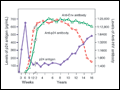 | Figure 189-27 Relationship between antigenemia and the development of antibodies to HIV. Antibodies to HIV proteins are generally seen 6–12 weeks following infection and 3–6 weeks after the development of plasma viremia. Late in the course of illness, antibody levels to p24 decline, generally in association with a rising titer of p24... |
view large |
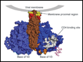 | Figure 189-28 Known targets of neutralizing antibodies against HIV-1. Group-specific antibody-binding regions include the membrane proximal region and the CD4 binding site. Type-specific antibody-binding regions include V2 and V3 loops. (Adapted from DR Burton et al:Nat Immunol 5:233, 2004.) |
view large |
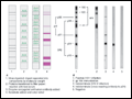 | Figure 189-29 Western blot assay for detection of antibodies to HIV. A . Schematic representation of how a Western blot is performed. B. Examples of patterns of Western blot reactivity. In each instance the Western blot... |
view large |
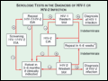 | Figure 189-30 Algorithm for the use of serologic tests in the diagnosis of HIV-1 or HIV-2 infection.*Stable indeterminate Western blot 4–6 weeks later makes HIV infection unlikely. However, it should be repeated twice at 3-month intervals to rule out HIV infection. Alternatively, one may test for HIV-1 p24 antigen or HIV RNA. EIA, enzyme... |
view large |
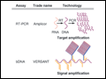 | Figure 189-31 Comparison of RT-PCR and bDNA assays. A. Schematic representation of reverse transcriptase–polymerase chain reaction (RT-PCR) and bDNA assays. See text for detailed description. B. Scatter plot of... |
view large |
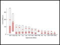 | Figure 189-32 Relationship between CD4+ T cell counts and the development of opportunistic diseases. Boxplot of the median (line inside the box), first quartile (bottom of the box), third quartile (top of the box), and mean (asterisk) CD4+ lymphocyte count at the time of the development of opportunistic disease.... |
view large |
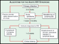 | Figure 189-33 The acute HIV syndrome. See text for detailed description. (Adapted from G Pantaleo et al: N Engl J Med 328:327, 1993. Copyright 1993 Massachusetts Medical Society. All rights reserved.) |
view large |
 | Figure 189-34 A. Decrease in the incidence of opportunistic infections and Kaposi's sarcoma in HIV-infected individuals with CD4+ T cell counts <100/μL from 1992 through 1998. [Adapted and updated from FJ Palella et al: N Engl J Med 338:853, 1998, and JE Kaplan et al: Clin Infect Dis... |
view large |
 | Figure 189-35 Various oral lesions in HIV-infected individuals. A. Thrush. B. Hairy leukoplakia. C. Aphthous ulcer. D. Kaposi's... |
view large |
 | Figure 189-36 Barium swallow of a patient with Candida esophagitis. The flow of barium along the mucosal surface is grossly irregular. |
view large |
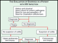 | Figure 189-37 Algorithm for the evaluation of diarrhea in a patient with HIV infection. HIV-associated enteropathy is a diagnosis of exclusion and can be made only after other, generally treatable, forms of diarrheal illness have been ruled out. |
view large |
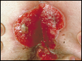 | Figure 189-38 Severe, erosive perirectal herpes simplex in a patient with AIDS. |
view large |
 | Figure 189-39 Characteristics of lipodystrophy. A. Truncal obesity and buffalo hump. B. Facial wasting. C. Accumulation of intraabdominal fat on CT scan. |
view large |
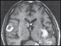
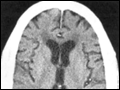 | Figure 189-40 AIDS dementia complex. Postcontrast CT scan through the lateral ventricles of a 47-year-old man with AIDS, altered mental status, and dementia. The lateral and third ventricles and the cerebral sulci are abnormally prominent. Mild white matter hypodensity is also seen adjacent to the frontal horns of the lateral ventricles. |
view large |
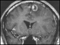 | Figure 189-41 Central nervous system toxoplasmosis. A coronal postcontrast T1-weighted MRI scan demonstrates a peripheral enhancing lesion in the left frontal lobe, associated with an eccentric nodular area of enhancement (arrow); this so-called eccentric target sign is typical of toxoplasmosis. |
view large |
 | Figure 189-42 Kaposi's sarcoma in three patients with AIDS demonstrating (A) periorbital edema and bruising; (B) classic truncal distribution of lesions; and (C) upper extremity l... |
view large |
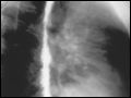
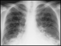 | Figure 189-43 Chest x-ray of a patient with AIDS and pulmonary Kaposi's sarcoma. The characteristic findings include dense bilateral lower lobe infiltrates obscuring the heart borders and pleural effusions. |
view large |
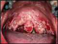
 | Figure 189-44 Immunoblastic lymphoma involving the hard palate of a patient with AIDS. |
view large |
 | Figure 189-45 Central nervous system lymphoma. Postcontrast T1-weighted MRI scan in a patient with AIDS, an altered mental status, and hemiparesis. Multiple enhancing lesions, some ring-enhancing, are present. The left sylvian lesion shows gyral and subcortical enhancement, and the lesions in the caudate and splenium ( |
view large |
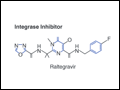 | Figure 189-46 Molecular structures of antiretroviral agents. |
view large |
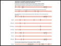 | Figure 189-47 Amino acid substitutions conferring resistance to antiretroviral drugs. For each amino acid residue, the letter above the bar indicates the amino acid associated with wild-type virus and the letter(s) below indicate the substitution(s) that confer viral resistance. The number shows the position of the mutation in the protein.... |
view large |