

PART 8: Infectious Diseases
SECTION 10 Diseases Caused by Rickettsiae, Mycoplasmas, and Chlamydiae
174 Rickettsial Diseases
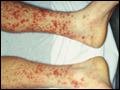 | Figure 174-1 Top: Petechial lesions of Rocky Mountain spotted fever on the lower legs and soles of a young, previously healthy patient. Bottom: Close-up of lesions from the same patient. (Photos courtesy of Dr. Lindsey Baden; with... |
view large |
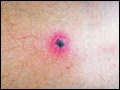 | Figure 174-2 Eschar at the site of the mite bite in a patient with rickettsialpox. (Reprinted from A Krusell et al: Emerg Infect Dis 8:727, 2002. Photo obtained by Dr. Kenneth Kaye.) |
view large |
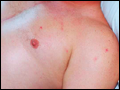 | Figure 174-3 Top: Papulovesicular lesions on the trunk of the patient with rickettsialpox shown in |
view large |
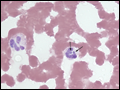 | Figure 174-4 Peripheral blood smear from a patient with human granulocytotropic anaplasmosis. A neutrophil contains two morulae (vacuoles filled with A. phagocytophilum). (Photo courtesy of Dr. J. Stephen Dumler.) |
view large |