

PART 8: Infectious Diseases
SECTION 9 Spirochetal Diseases
171 Leptospirosis
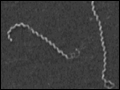 | Figure 171-1 Transmission electron micrograph of Leptospira interrogans serovar Icterohaemorrhagiae. |
view large |
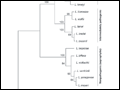 | Figure 171-2 Differentiation of pathogenic, intermediately pathogenic, and nonpathogenic (saprophytic) Leptospira species based on molecular phylogenetic analysis using the 16S rRNA gene. Scale bar indicates rate of nucleotide substitution per base pair. |
view large |
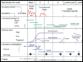 | Figure 171-3 Biphasic nature of leptospirosis and relevant investigations at different stages of disease. Specimens 1 and 2 for serology are acute-phase serum samples; specimen 3 is a convalescent-phase serum sample that may facilitate detection of a delayed immune response; and specimens 4 and 5 are follow-up serum samples that... |
view large |
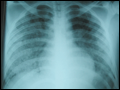 | Figure 171-4 Severe pulmonary hemorrhage in leptospirosis. Left: Chest x-ray. Right: Gross appearance of right lower lobe of lung at autopsy. This patient, a 15-year-old in the... |
view large |