

PART 8: Infectious Diseases
SECTION 6 Diseases Caused by Gram-Negative Bacteria
143 Meningococcal Infections
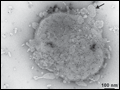 | Figure 143-1 Electron micrograph of Neisseria meningitidis. Black dots are gold-labeled polyclonal antibodies binding surface opacity proteins. Blebs of outer membrane can be seen being released from the bacterial surface (see arrow). (Photo courtesy of D. Ferguson, Oxford University.) |
view large |
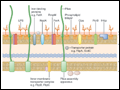 | Figure 143-2 Cross-section through surface structures of Neisseria meningitidis. (Reprinted with permission from Sadarangani and Pollard, 2010.) |
view large |
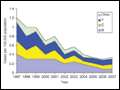 | Figure 143-3 Meningococcal disease in the United States over time. (Adapted from ABC Surveillance data, Centers for Disease Control and Prevention; www.cdc.gov.) |
view large |
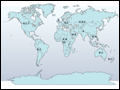 | Figure 143-4 Global distribution of meningococcal serogroups, 1999–2009. |
view large |
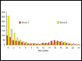 | Figure 143-5 Age distribution of serogroups B and C meningococcal disease in England and Wales, 1998/1999. (Health Protection Agency, UK; www.hpa.org.uk.) |
view large |
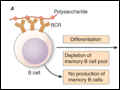 | Figure 143-6 A. Polysaccharides from the encapsulated bacteria that cause disease in early childhood stimulate B cells by cross-linking the BCR and driving the production of immunoglobulins. There is no production of memory B cells, and the B cell pool may be depleted by this process such that subsequent immune... |
view large |
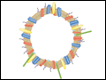 | Figure 143-7 Illustration of meningococcal outer-membrane vesicle containing outer-membrane structures. |
view large |