

PART 8: Infectious Diseases
SECTION 5 Diseases Caused by Gram-Positive Bacteria
134 Pneumococcal Infections
 | Figure 134-1 Pneumococci growing on blood agar, illustrating α hemolysis and optochin sensitivity (zone around optochin disk). Inset: Gram's stain, illustrating gram-positive diplococci. (Photographs courtesy of Paul Turner, Shoklo Malaria Research Unit, Thailand.) |
view large |
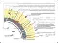 | Figure 134-2 Schematic diagram of the pneumococcal cell surface, with key antigens and their roles highlighted. |
view large |
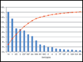 | Figure 134-3 Meta-analysis of available global serotype data, adjusted for regional disease incidence. The red line shows cumulative incidence, as indicated on the right-hand Y axis. (Source: Global Serotype Project Report for the Pneumococcal Advance Market Commitment Target Product Profile; available at |
view large |
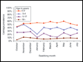 | Figure 134-4 Prevalence of pneumococcal carriage in adults and children resident in the United Kingdom who had nasopharyngeal swabs collected monthly for 10 months (no seasonal trend; t test trend, >.05). (Data adapted from D Goldblatt et al, 2005.) |
view large |
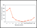 | Figure 134-5 Rates of invasive pneumococcal disease before the introduction of pneumococcal conjugate vaccine, by age group: United States, 1998. [Source: CDC, Active Bacterial Core Surveillance/Emerging Infectious Program Network, 2000. Data adapted from MMWR 49(RR-9), 2000.] |
view large |
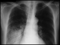 | Figure 134-6 Chest radiograph depicting classic lobar pneumonia in the right lower lobe of an elderly patient's lung. |
view large |
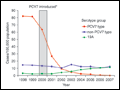 | Figure 134-7 Changes in invasive pneumococcal disease (IPD) incidence, by serotype group, among children <5 years old (top) and adults >65 years old (bottom), 1998–2007. *7-Valent pneumococcal conjugate vaccine (PCV7) was introduced in the United States for routine... |
view large |